Download
1 / 38
430 likes | 655 Vues
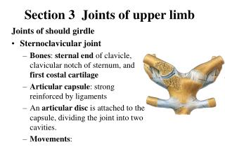
JOINTS OF THE UPPER LIMB. Connection of the upper limb Connection of the shoulder girdle (juncturae ossium cinguli extremitatis superioris) 1. Articulatio sternoclavicularis (compoud joint) Articular surfaces: incisura clavicularis sterni and facies articularis sternalis of clavicula .

E N D
Connection of the upper limb Connection of the shoulder girdle (juncturae ossium cinguli extremitatis superioris) 1. Articulatio sternoclavicularis (compoud joint) Articular surfaces: incisura clavicularis sterni and facies articularis sternalis of clavicula. Articular capsule: is stiff and is attached to margines of articular surfaces Auxiliary features: discus articularis, lig. sternoclaviculare anterius and posterius., lig. interclaviculare, lig. costoclaviculare Type of joint: ball–and–socket joint with movements to all directions but movement are limited, a component of movements of scapula and shoulder joint. 2. Articulatio acromioclavicularis(usually a compound joint) Articular surfaces: facies articularisclavicularis of acromionand facies articularis acromialis of clavicula. Articular capsule: is attached to margines of articular surfaces Auxiliary features: often is present discus articularis, lig. acromioclaviculare, lig. coracoclaviculare Type of joint: ball–and–socket joint with movements to all directions but movement are limited, a component of movements of scapula and shoulder joint.
3. Ligaments of the scapula Lig. transversum scapulae Lig. coracoacromiale – between processus coracoideus and akromion of scapula. Together with both bone processus forms fornix humeri. Abduction of shoulder joint is always associated with movements of scapula!
Connections of the free part of the upper limb (juncturae ossium extremitatis superioris) 1. Shoulder joint(articulatio humeri) Articular surfaces: caput humeri and cavitas glenoidalis of scapula Articular capsule: is attached to margines of cavitas glenoidalis, reaches collum anatomicum of humerus, on the medial side of humerus runs distally (folds of capsule for abduction). Ventrally the synovial layer of articular capsule covers tendon of long head of m. biceps brachii and forms – vagina synovialis intertubercularis. Auxiliary features: labrum glenoidale,ligg. glenohumeralia, lig. coracohumerale. Articular capsule is reinforced by tendons of muscles (m. subscapularis, m. supraspinatus, m. infraspinatus, m. teres minor). Type of joint: ball-and-socket, movements are possible to all directions (three degrees of freedom of movements).
2. Elbow joint(articulario cubiti) – compound joint Articulatio humeroradialis Articular surfaces: capitulum humeri and fovea capitis of radius Articulatio humeroulnaris Articular surfaces: trochlea humeri and incisura trochlearis of ulna Articulatio radioulnaris Articular surfaces: circumferentia articularis capitis radii and incisura radialis ulnae Articular capsule: both epicondyli of humerus are free (they serve for attachment of muscles), all fossae of distal end of humerus are located intracapsularly, on the radius runs to the collum radii – recessus sacciformis. Auxiliary features: lig. anulare radii, lig. collaterale radiale and lig. collaterale ulnare. Type of joint: Articulatio humeroradialis is ball-and-socket joint, articulatio humeroulnaris is a hinge joint and articulatio radioulnaris proximalis is a trochoid joint. Movements here are limited by position of olecranon ulnae in fossa olecrani. Is possible only flexion and extension, rotation (inner-pronation) and external rotation (supination).
Connections of antebrachium(juncturae radioulnares) Articulatio radioulnaris proximalis, articulatio radioulnaris distalis and membrana interossea natebrachii. A. Articulatio radioulnaris distalis Articular surfaces: caput ulnae and incisura ulnaris radii Articular capsule: is thinandfree Auxiliary features: together witharticulatio radiocarpea Type of joint: trochoid joint – rotation. B. Membrana interossea antebrachii – stiff membrane attached to margo interosseus of radius and ulna. It serves for attachment of some muscles of forarm, it limits external rotation.
4. Joints of the hand(articulationes manus) A. Articulatio radiocarpea Articular surfaces: facies articularis carpea radii and os scaphoideum, os lunatum and os triquetrum. Articular capsule: together with articulatio mediocarpea Auxiliary features: discus articularis, ulna is separated from carpal bones by this discus. Ligaments shares with articulatio mediocarpea. Type of joint: ellipsoidal, movements together with articulatio mediocarpea. B. Midcarpal joint (articulatio mediocarpea) – connection between proximal and distal row of carpal bones Articular surfaces : laterally – trapezium (os trapezium) and trapezoideum (os trapezoideum) form the articular fossa and scaphoideum (os scaphoideum) forms an articular head, medially – scaphoid (os scaphoideum), lunate and triquetrum (os lunatum and os triquetrum) form an articular fossa and articular head is formed by capitate and hamate (os capitatum and os hamatum). Joint has an S-shaped joint space. Articular capsule : shares with radiocarpal joint (articulatio radiocarpea) Additional features: dorsal and palmar radiocarpal ligaments (lig. radiocarpeum dorsale and palmare), palmar ulnocarpal ligament (lig. ulnocarpeum palmare), radiate carpal ligament (lig. carpi radiatum) runs from palmar surface of capitate (os capitatum) to the neighbour carpal bones.Dorsal, palmar and interosseous intercarpal ligaments (ligg. intercarpea dorsalia, palmaria and interossea) join together carpal bones. Type of joint : ellipsoid joint, movements shares together with midcarpal joint (articulatio mediocarpea) – palmar and dorsal flexion, radial and ulnar deviation and rotary movement .
C. Articulatio ossis pisiformis Articular surfaces: connection between os pisiforme and os triquetrum. Articular capsule: is attached to margins of the articular surfaces. Auxiliary features: articular capsule is reinforced by lig. pisohamatum and lig. pisometacarpeum. D. Articulatio carpometacarpea pollicis Articular surfaces: connection between os trapezium and basis of the I. metakarpal bone. Articular capsule: is relatively free and it is attached to margins of the articular surfaces. Type of the joint: saddle; movements – abduction and adduction of the thumb, oposition and reposition. Thunb is the most movable finger. E. Articulationes carpometacarpeae II. – V. Articular surfaces: distal row of carpal bones joins to bases of the II. – V. metakarpal bones. Connection between sides of bases of metacarpal bones. Articular capsule: is attached to margins of the articular surfaces. Auxiliary features: Ligg. metacarpea palmaria,dorsalia and interossea and between bases of metacarpal bones ligg. metacarpea palmaria, dorsalia and interossea. Type of the joint: amphiartrosis (almost immobile joint).
F. Articulationes metacarpophalangeae Articular surfaces: caput of metacarpals and bases of proximal phalanges Articular capsule: is attached to margins of the articular surfaces. Auxiliary features: connective plates increase articular pits – laminae fibrocartilagineae palmares and ligg. collateralia. Metacarpophalangeal joint has in lamina fibrocartilaginea two small sesamoid bones. Palmar side of caput the II. – V. metakaral bones are joined by lig. metacarpeum transversum profundum. Type of the joint: ellipsoidal, with possibility of flexion, extension, abduction and adduction. G. Articulationes interphalangeae manus Articular surfaces : trochlea phalangis of the proximal phalanx, basis of distal phalanx. Articular capsule : is attached to margins of the articular surfaces. Auxiliary features : connective plate increases articular pit – lamina fibrocartilaginea palmaris. Articular capsule is reinforced by ligg. collateralia. Auxiliary features : hinge joint, movements – flexion and extension, distal phalanx with possibility of hyperextension.
Connection of the lower limb (juncturae ossium extremitatis inferioris) includes connection of pelvic girdle and free part of lower limb. Connection of pelvic girdle (juncturae ossium cinguli extremitatis inferioris) they have relation to pelvis, which arises by joining of two pelvic bones and dorsally with sacral bone. 1. Articulatio sacroiliaca (sacroiliac joint) Articular surfaces: facies auricularis of sacral and hip bones. Articular capsule: is taut and is attached to the articular surfaces. Auxiliary features: capsule is strengthened byligg. sacroiliaca ventralia and dorsalia,ligg.sacroiliacainterossea are located between tuberositas sacralis and tuberositas iliaca. Auxiliary features: amphiartrosis (with minimal movements).
2. Symphysis pubica Is formed by cartilagenous discus interpubicus which connects both pubic bones. Symphysis pubica is 4,5 – 5 cm in hight. There is on the upper margin of symphysis lig. pubicum superius, under it very strong lig. arcuatum pubis. 3. Membrana obturatoria Is a stiff membrane which closes foramen obturatum; it serves as attachment for mm. obturatorii.
4. Ligaments in the pelvic region Lig. sacrospinale (fan out to the lateral margin of the sacral bone from the spina ischiadica). Lig. sacrotuberale (fan out to the lateral margin of the sacral bone from the tuber ischiadicum). Incisura ischiadica major is converted by course of lig. sacrospinale into foramen ischiadicum majus (greater sciatic foramen). This foramen is divided by m. piriformis into foramen suprapiriforme and foramen infrapiriforme (content – nerves and vessels to gluteal muscles). Foramen ischiadicum minus (lesser sciatic foramen) is limited by ligamentum sacrotuberale and sacrospinale and incisura ischiadica minor. Through this opening run m. obturatorius internus and nerves and vessels to external genital organs). Lig. iliolumbale passes from processus costarius of 4. and 5. lumbar vertebrae to crista iliaca (iliac crest).
5. Pelvis The bony pelvis consists of two hip bones, ventrally are joined by cartilaginous symphysis pubis and dorsally by os sacrum and os coccygis. Aditus pelvis (the pelvic inlet) is bordered by linea terminalis which separatespelvis major, located above this linea (a part of the abdominal cavity) and pelvis minor (its content – a part of genital and urinary systems). Exitus pelvis (the pelvic outlet) is the region between the subpubic angle, tubera ischiadica and os coccygis. Pelvis minor is an important childbirth way in female and it has an great intersexual differences. Male pelvis is higher and narrower, incisura ischiadica major has shape like letter J, longitudinally oriented foramen obturatum and angulus subpubicus. Female pelvis is lower and wider than in the male. Foramen obturatum is transversely directed and female pelvis has arcus pubicus, incisura ischiadica major has shape like broad V letter. The absolute diameters are longer in pelvis of males.