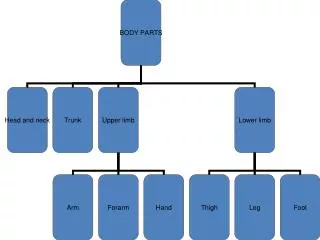
Section 3 Joints of upper limb
460 likes | 1.12k Vues
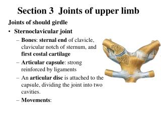
Section 3 Joints of upper limb. Joints of should girdle Sternoclavicular joint Bones : sternal end of clavicle, clavicular notch of sternum, and first costal cartilage Articular capsule : strong reinforced by ligaments

Section 3 Joints of upper limb
E N D
Presentation Transcript
Section 3 Joints of upper limb Joints of should girdle • Sternoclavicular joint • Bones: sternal end of clavicle, clavicular notch of sternum, and first costal cartilage • Articular capsule: strong reinforced by ligaments • An articular disc is attached to the capsule, dividing the joint into two cavities. • Movements:
coracoacromial ligament acromion coranoid process • Acromioclavicular joint • Bones: acromion and acromial end of clavicle • Movement: rotation of scapula on clavicle • Coracoacromial arch formed by coracoacromial ligament, coranoid process, and acromion, that prevents the shoulder joint from superior dislocation
Joints of free upper limb ★Shoulder joint (ball and socket) • Bones: head of humerus and glenoid cavity of scapula • Capsule: • Thin and lax, especially lower part • Tendon of long head of biceps brachii passes though the cavity
Accessory structures • Glenoid labrum:fibrocartilaginous ring on periphery of glenoid cavity • Coracohumeral ligament:runs from coracoid process to greater tubercle • Movements: flexion-extension, adduction-abduction, medial and lateral rotation, circumduction
★Elbow joint • Bones: lower end of humerus, upper ends of radius and ulna • Humeroulnar joint : formed by trochlear of humerus and troclear notch (hinge) • Humeroradial joint: formed by capitulum of humerus and head of radius (ball and socket) • Proximal radioulnar joint: formed by articular circumference of radius and radial notch of ulna • Capsule: thin and lax anteriorly and posteriorly, strongly thickened on either side by collateral ligaments
Ligaments: • Radial collacteral ligament: attached to lateral epicondyle and annular ligament of radius • Ulnar collacteral ligament: attached to medial epicondyle to medial border of trochlear notch • Annular ligament of radius: attached to anterior and posterior margins of radial notch of ulna, surrounds the head of radius • Movements: flexion and extension, pronation and supination
Joints between radius and ulna • Proximal radioulnar joint • Distal radioulnar joint: formed by head of ulna, ulnar notch of radius and an articular disc • Interosseous membrane of forearm: a fibrous membrane between the shaft of radius and ulna
Joints of hand ★Radiocarpal joint(ellipsoid) • Bones • Carpal articular surface of radius and articular disc below the ulna • Proximal row of carpal: scaphoid, lunate, and triquetrum, but not pisiform • Capsule: lax and strengthened by surrounding ligament • Movements: flexion, extension, adduction, abduction, and circumduction
Intercarpal joints • Carpometacarpal joints: Carpometacarpal joint of thumb(saddle) • Bones: trapezium and base of first metacarpal • Movement: flexion, extension, adduction, abduction, and opposition • Intermetacarpal joints • Metacarpophalangeal joints • Interphalangeal joints
Section 4 Joints of Lower limb Joints of pelvic girdle Sacroiliac joint • Bones: auricular surface of sacrum and ilium • Capsule: very tight and strengthened by ligaments
★Sacrotuberous ligament:runs from lateral margins of sacrum and coccyx to the inner margin of ischial tuberosity ★Sacrospinous ligament:runs from ischial spine to lateral margins of sacrum and coccyx • These two ligaments convert the sciatic notches the greaterandlesser sciatic foramina
Sacrospinous ligament Greater sciatic foramen Sacrotuberous ligament Lesser sciatic foramen
Pubic symphysis Articulation: symphysial surface and interpubic disc (fibrocartilage) Ligaments: superior pubic ligament and arcuate pubic ligament Obturator membrane Obturator canal
Obturator canal Obturator membrane
Bony pelvis • Composition: formed by paired hip bones, sacrum, coccyx, and their articulations • In anatomical position, anterior superior iliac spines and pubic tubercles on same vertical plane, while the tip of coccyx and superior border of pubic symphysis on same horizontal plane • Terminal line: formed by promontory of sacrum, arcuate line, pectin of pubis, pubic tubercle, upper border of pubic symphysis • Two portions: a greater pelvis and a lesser pelvis
Lesser pelvis • pelvic inlet(terminal line): • Pelvic outlet : formed by tip of coccyx, sacrotuberous ligament, ischial tuberosity, ramus of ischium, inferior ramus of pubic, symphysis • Pelvic cavity • Pubic arch, subpubic angle
Main difference between male and femal pelvis Male Female Pelvic inlet Pelvic outet Pelvic cavity Pubic arch 90~1000 70~750
Joints of free lower limb ★Hip joint • Bones: acetabulum and femoral head • Articular capsule attachments • Above: margins of acetabulum and transverse acetebular ligament • Below: in front to intertrochanteric line; behind, to the neck of femur above 1cm above the intertrochanteric crest
Acetabulum labrum Ligament of head of femur Transverse acetebular lig. • Accessory structures • Acetabulum labrum, transverse acetebular ligament • Ligaments • Iliofemoral lig. • Ligament of head of femur • Pubofemoral lig. • Ischiofemoral ligament • Zona orbicularis • Movement: flexion-extention, adduction-abduction medial and lateral rotation, circumduction
Pubofemoral lig. Iliofemoral lig. Ischiofemoral lig. Zona orbicularis
★ Knee joint • Bones: lower end of femur, upper end of tibia and patella • Articular capsule: superapatellar bursa, deep infrapatellar bursa, ala folds
Fibular collateral lig. Patellar lig. Tibial collateral lig. • Accessory structures • Extra-ligaments • Patellar lig. • Fibular collateral lig. • Tibial collateral lig. • Oblique popliteal ligament
- Intra-ligaments • Anterior cruciate ligament • Posterior cruciateligament
Medial meniscus(C-shaped) • lateral meniscus(O-shaped) • Movements: flexion and extension; flexed knee joint may be passively rotated Medial lateral
Tibiofibular syndesmosis • Tibiofibular joint • Crural interosseous membrane • Anterior and posterior tibiofibular ligaments
Joint of foot Talocrural joint (ankle joint) • Bones: lower ends of tibia and fibula, talus • Articular capsule: thin and lax in front and behind, and supported on each side by strong collateral ligaments
Ligments • Medial lig. • Lateral lig. • Movements: dosiflexion (extension) and plantar flexion (flexion); when the ankle joint is fully plantar flexed, small amounts of abduction, and adduction are possible(wrench)
Intertarsal joints • Tarsometatarsal joints • Intermetatarsal joints • Metatarsophalangeal joints • Interphalangeal joints
Arches of foot • Medial longitudinal arch: formed by calcaneus, navicular, three cuneiforms and first to third metatarsal bones, head of talus is the keystone of this arch
Lateral longitudinal arch: formed by calcaneus, cuboid, fourth and fifth metatarsals; cuboid is the keystone of this arch
Tranverse arch: formed by cuboid, three cuniforms and all metatarsals; the intermediate cuneiform is the keystone of this arch • Function: give to foot strength stability and resilience; protect plantar vessels and nerves
Normal arch Flatfoot
Section 5 Joints of skull • Continuous joints: sutures, synchondrosis or synosteosis
Temporomandibular joint • Aticulating surfaces • Mandibular fossa and articular tubercle, above • Head of mandible, below • Capsule: thin and lax in front and behind; strengthened by the lateral ligament • Articular disc: separates surfaces, forming upper and lower compartments within joint • Movement: mandible may be elevated or depressed, protruded or retracted; rotation may also occurs as in chewing( a slight amount of side to side movement is also permitted)