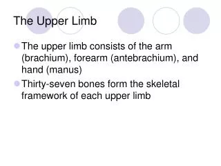
The upper limb
The upper limb. 山东大学医学院 解剖教研室 李振华. The muscles of back. Superficial group Trapezius 斜方肌 Latissimus dorsi 背阔肌 Levator scapulae 肩胛提肌 Rhomboideus 菱形肌 Deep group Erector spinae 竖脊肌 Splenius 夹肌 Thoracolumbar fascia 胸腰筋膜. The muscles of thorax. Extrinsic muscles


The upper limb
E N D
Presentation Transcript
The upper limb 山东大学医学院 解剖教研室 李振华
The muscles of back Superficial group • Trapezius斜方肌 • Latissimus dorsi背阔肌 • Levator scapulae 肩胛提肌 • Rhomboideus菱形肌 Deep group • Erector spinae 竖脊肌 • Splenius 夹肌 • Thoracolumbar fascia 胸腰筋膜
The muscles of thorax Extrinsic muscles • Pectoralis major胸大肌 • Pectoralis minor胸小肌 • Serratus anterior 前锯肌 Intrinsic muscles • Intercostales externi肋间外肌 • Intercostales interni肋间内肌 • Intercostales intimi 肋间最内肌
Major muscles of the trunk Trapezius • Origin: superior nuchal line, external occipital protuberance, ligamentum nuchae and spinous processes of seventh cervical and all thoracic vertebrae • Insertion: lateral third of clavicle, acromion, and spine of scapulartery • Acton: upper fibers elevate scapula, lower fibers depress scapula; if scapula is fixed, one side acting along, draws head toward the same side, and turn face to opposite side; both sides together, draw head directly backward
Latissimus dorsi • Origin: spinous processes of lower six thoracic and all lumbar vertebrae, median sacral crest, and posterior part of iliac crest. • Insertion: floor of intertubercular groove of humerus. • Action: trunk fixed, extends, adducts and medially rotates arm ; arm fixed, elevates trunk.
Erector spinae • Position: fills the vertebral groove on each side of the spine • Action: extends vertebral column (draw head backwar)
Pectoralis major • Origin: medial half of clavicle,sternum,1th-6th costal cartilages. • Insertion: crest of greater tubercle of humerus. • Action: flexes, adducts and rotates arm medially; arm fixed, elevates trunk; elevates ribs 1-6,aidding in forced inspiration.
The Muscles of Upper Limb Muscles of shoulder • Deltoid • supraspinatus • Infraspinatus • Teres minor • Teres major • subscapularis
Muscles of arm • Antererior group • Biceps brachii 肱二头肌 • Coracobrachialis喙肱肌 • Brachialis 肱肌 • Posterior group – triceps brachii肱三头肌
Muscles of forearm • Superficial layer • Brachioradialis 肱桡肌 • Pronator teres 旋前圆肌 • Flexor carpi radialis 桡侧腕屈肌 • Palmaris longus 掌长肌 • Flexor carpi ulnaris 尺侧腕屈肌
Third layer • Flexor digitorum profundus 指深屈肌 • Flexor pollicis longus 拇长屈肌 • Fourth layer- pronator quadratus旋前方肌 Action: flex radiocarpal joint and fingers, pronate forearm
Posterior group (10) • Superficial layer (5) • Extensor carpi radialis longus 桡侧腕长伸肌 • Extensor carpi radialis brevis 桡侧腕短伸肌 • Extensor digitorum 指伸肌 • Extensor digiti minimi 小指伸肌 • Extensor carpi ulnaris 尺侧腕屈肌
Deep layer (5) • Supinator旋后肌 • Abductor pollicis longus拇长展肌 • Extensor pollicis brevis拇短伸肌 • Extensor pollicis longus拇长伸肌 • Extensor indicis 示指伸肌 • Action: extend radiocapral joint and fingers, and supinate forearm
Muscles of hand • Lateral group-thenar 鱼际(4) • Abductor pollicis brevis拇短展肌 • Flexor pollicis brevis 拇短屈肌 • Opponens pollicis 拇对掌肌 • Adductor pollicis 拇收肌 • Action: flex, abduct, adduct and oppose thumb • Medial group-hypothenar (3) • Abductor digiti minimi 小指展肌 • Flexor digiti minimi brevis小指短屈肌 • Opponens digiti minimi小指对掌肌 • Action: flex, abduct , and oppose little finger
Intermedial group • Lumbricales 蚓状肌(4)-flex fingers at MP joints; extend fingers at IP joints • Palmar interossei 骨间掌侧肌(3)- adduct fingers towards middle finger at MP joints • Dorsal interossei 骨间背侧肌(3)-abduct fingers away from middle finger at MP joints
Major muscles of upper limb Deltoid • Origin: lateral third of clavicle, acromion, and spine of scapula • Insertion: deltoid tuberosity of humerus • Action: abducts,flexes and medically rotates, extends, and laterally rotates arm
Teres major • Origin: dorsal surface of inferior angle of scapula • Insertion: crest of lesser tubercle of humerus • Action: medially rotates and adducts arm
Biceps brachii • Origin: long head, supraglenoid tubercle; short head, coracoid process • Insertion: radical tuberosity • Action: supinator of forearm, flexor of elbow joint, weak flexor of should joint Pronator teres • Origin: medical epicondyle of humerus and deep fascia of forearm • Insertion: middle of lateral surface of radius • Action: pronation of forearm and flexion of elbow
Triceps brachii • Origin: long head, infraglenoid tubercle; lateral head, above groove for radical n., medical head, below groove for radical n. • Insertion: olecranon of ulna • Action: extends elbow joint), long head can extend and adduct shoulder joint
Supinator • Origin: lateral epicondyle of humerus and upper part of lateral border of ulna • Insertion: upper third of anterior surface of radius • Action: supination of forearm
Arteries of upper limb Axillary artery • Continuation of subclavian artery at lateral border of first rib • Becomes brachial artery at lower border of teres major • Divided into three parts by overlying pectoralis minor • First portion, above muscle-gives rise to thoracoacromial a. 胸肩峰动脉 • Second portion, behind muscle-gives rise to lateral thoracic a. 胸外侧动脉 • Third portion, below muscle-gives rise to subscapular a. 肩胛下动脉, anterior and posterior humeral circumflex a. 旋肱前、后动脉; the former then divides into throcodorsal a. 胸背动脉and circumflex scapular a. 旋肩胛动脉
Brachial artery • Continuation of axillary artery • Divides into radial and ulnar arteries at level of neck of radius • Branches • Deep brachial a. 肱深动脉-accompanies with radial nerve • Superior ulnar collaeral a. 尺侧上副动脉-accompanies with ulnar nerve • Inferior ulnar collateral a.尺侧下副动脉
Radial artery and branches • Radial recurrent a. 桡侧返动脉 • Superfical palmar branch 掌浅支 • Principal artery of thumb 拇主要动脉 Ulnar artery and branches • Ulnar recurrent a. 尺侧返动脉 • Common interosseous artery 骨间总动脉 • Anterior interossous a. 骨间前动脉 • Posterior interosseous a. 骨间后动脉 • Deep palmar branch 掌深支
Superficial palmar arch 掌浅弓 • Formed by ulnar artery and superficial palmar branch of radial artery • Curve of arch lies across the palm, level with the distal border of fully extended thumb • Gives rise to three common palmar digital arteries each then divides into two proper palmar digital arteries
Deep palmar arch 掌深弓 • Formed by radial artery and deep palmar branch of ulnar artery • Curve of arch lies across upper part of palmar at level with proximal border of extended thumb • Gives rise to three palmar metacarpal arteries
Veins of the upper limb Deep veins: accompany the arteries of the same region and bear similar names Superficial veins • Cephalic vein 头静脉 • Arises from the lateral side of the dorsal venous rete of hand • Ascends on radial side of the forearm to the elbow and then in the lateral side of biceps brachii furrow, continues up the arm in the deltopectoral groove and then to the infraclavicular fossa, where it pierces clavipectoral fascia to drain into axillary vein
Basilic vein贵要静脉 • Arises from the medial side of the dorsal venous rete of hand • Ascends on the ulnar side of forearm to the elbow and then in the medial bicepital brachii furrow to middle of the arm where it pierces the deep fascia and joins the brachial vein or axillary vein • Median cubital vein 肘正中静脉 links cephalic vein and basilic vein in the cubital fossa. It is a frequent site for venipuncture to remove a sample of blood or add fluid to the blood
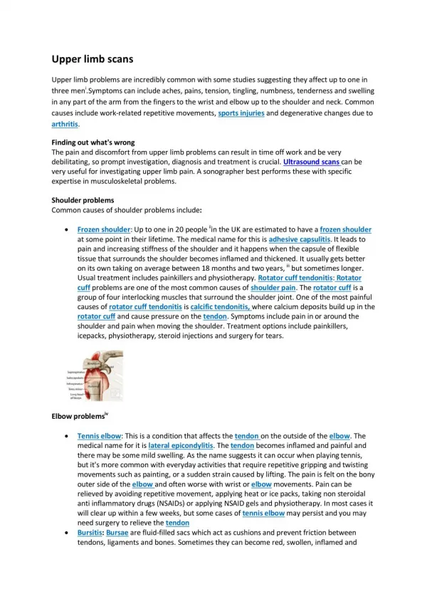
The lymphatic drainage of upper limb Lymphatic vessels • Superficial-follow the superficial veins, drain into supratrochlear and axillary lymph nodes • Deep-accompany main vessels, end in axillary lymph nodes lymph nodes • Cubital lymph node: lies above medial epicondyle of humerus • Axillary lymph node-arranged in five groups
Axillary lymph nodes腋淋巴结-arranged in five groups • Lateral lymph nodes 外侧淋巴结- lie around the distal end of axillary vein , receiving drainage from the arm, forearm, and hand • Pectoral lymph nodes 胸肌淋巴结-lie along lateral thoracic vessels, receive afferents from anterior thoracic wall including central and lateral portion of mamma • Subscapular lymph node 肩胛下淋巴结-along subscapular vessels, receive lymph from nape and scapular region • Efferents above three groups pass to central lymph node
Central lymph node中央淋巴结-lie in fat of axillary fossa, receive drainage from all the above nodes, efferents pass to apical lymph node • Apical lymph node 尖淋巴结 • Lie in the apex of the axilla, along the proximal end of axillary vessels • Receive drainage chiefly from central lymph node , upper portion of mamma • Efferents form subclavian trunk, the right subclavian trunk joints the right lymphatic duct; left usually drains directly into thoracic duct
Brachial plexus 臂丛 Formation: • Five roots: formed by anterior rami of C5-C8 and T1 spinal nerves, roots C5~C7give rise to long thoracic n.胸长神经 • Three trunks • The upper trunk is formed by the joining of root C4,C5,C6. • The middle trunk is the continuation of root C7. • The lower trunk is formed by the joining of root C8 and T1. • Six divisions: above clavicle, trunks form anterior and posterior divisions • Three cords: below clavicle, divisions form three cords that surround the second portion of axillary a.
Position: passes through the scalene fissure to posterosuperior of subclavian artery, then enters the axilla to form lateral, medial and posterior cords Main branches • Lateral cord • Musculocutaneous n. 肌皮神经 • Lateral root to median n. 正中神经外侧根 • Medial cord • Medial root to median n. 正中神经内侧根 • Ulnar n. 尺神经 • Medial brachial cutaneous n.臂内侧皮神经 • Medial antebrachial cutaneous n. 前臂内侧皮神经
Posterior cord • radial n. 桡神经 • axillary n. 腋神经 • thoracodorsal n. 胸背神经
Musculocutaneous肌皮神经 Distribution: Biceps brachii, brachalis and coracobrachialis ‘BBC nerve’; skin on anterior aspect of forearm
Median正中神经 • Distribution: Flexor of forearm except brachioradialis, flexor carpi ulnaris and ulnar half of flexor digitorum profundus, thenar except adductor pollicis, first two lumbricals; skin of thenar, central part of palm, palmar aspect of radial three and one-half fingers, including middle and distal fingers on dorsum • Injury: Apehand 猿手 produces sign of benediction, in which the index and middle fingers cannot be flexed and the thumb cannot be opposed
Ulnar nerve • Distribution: Flexor carpi ulnaris, ulnar half of flexor digitorum profundus, hypothenar muscles, interossei, 3rd and 4th lumbricals and adductor pollicis; skin of hypothenar, palmar surface of ulnar one and one-half fingers, ulnar half of dorsum of hand, posterior aspect of ulnar two and one-half fingers • Injury: clawhand
Radial桡神经 • Distribution: Extensor muscles of arm and forearm, brachioradialis; skin on back of arm, forearm, and radial side of dorsum of hand and radial two and one-half fingers • Injury: Wristdrop
Axillary 腋神经 • Distribution: Deltoid and teres minor muscle; skin over deltoid and upper posterior aspect of arm • Injury: result in deltoid andteres minor paralysis (loss of shoulser abdution and weel external rotation) with loss of sensation over the deltoid
Regional anatomy of upper limb 山东大学医学院 解剖教研室 李振华
Parts and regions • Shoulder region-junction of arm and trunk • Arm-between should and elbow • Elbow-bend of arm, joint between arm and forearm • Forearm-between elbow and hand • Hand
Surface anatomy • Shoulder region: acromion, spine of scapula, coracoid process, greater tubercle, anterior and posterior axillary folds • Arm-medial and lateral biceps brachii furrow, deltoid tuberosity • Elbow-medial and lateral epicondyles, head of radius, olecranon, tendon of biceps brachii • Forearm-between elbow and hand • Hand-styloid process, dorsal tubercle
Anatomical snuff box 鼻烟壶 • When the thumb is abducted and extended, a triangular hollow appears between the tendon of the extensor pollicis longs medially and the tendons of the extensor pollicis brevis and abductor pollicis longus laterally. • The floor of the snuff box is the scaphoid and trapezium bones and crossed by the radial a..
Carring angle 提携角 1650~1700
Mamma 乳房 Position • Lie in superficial fascia over the pectorals major and pectoral fascia • Extend from 3rd to 7th ribs vertically, and from parasternal line to midaxillary line transversally
Structures-contains skin, mammary glands and adipose tissue • Each brest has about 15~20 lobes of mammary gland • Each lobe radiates out from the nipple and has a lactiferous duct输乳管which opens separately on the summit of the nipple and possesses a dilated lactiferous sinuses输乳管窦just prior to its termination
Suspensory ligaments of breast乳房悬韧带(cooper’s ligaments )-strands of connective tissue runs between skin and deep fascia and serve to support the mammary glands
Axillary fossa 腋窝 • The axillary fossa is a pyramid-shaped space through which major neurovascular structures pass between the thorax and upper extremity
Boundaries • The apex is a triangular space limited by the first rib, the scapula, and the middle third of clavicle. • The base-skin and fascia of the axillary fossa