
BIOCHEMICAL TESTS FOR LIVER DISEASE:
Module 1. BIOCHEMICAL TESTS FOR LIVER DISEASE: There are three major groups of tests (all based on serum/plasma levels). 1. Enzymes those that indicate hepatocellular leakage (ALT, AST, GLDH) those that indicate cholestasis (ALP, GGT). 2. Tests of clearance/excretory ability bilirubin

BIOCHEMICAL TESTS FOR LIVER DISEASE:
E N D
Presentation Transcript
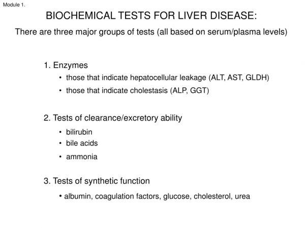
Module 1. BIOCHEMICAL TESTS FOR LIVER DISEASE: There are three major groups of tests (all based on serum/plasma levels) • 1. Enzymes • those that indicate hepatocellular leakage (ALT, AST, GLDH) • those that indicate cholestasis (ALP, GGT) • 2. Tests of clearance/excretory ability • bilirubin • bile acids • ammonia • 3. Tests of synthetic function • albumin, coagulation factors, glucose, cholesterol, urea
Module 1. BIOCHEMICAL TESTS FOR LIVER DISEASE: Let’s start with the enzymes… • 1. Enzymes • those that indicate hepatocellular leakage (ALT, AST, GLDH) • those that indicate cholestasis (ALP, GGT)
Module 1. These necrotic periacinar hepatocytes will be leaking ALT Of the hepatocellular leakage enzymes, the classic example is ALT • ALT leaks from hepatocyte in response to necrosis or sublethal cell injury • Relatively “liver specific” • Serum clearance half life: dog 2.5 days • Mild induction by corticosteroids (in some species, e.g. dog) and other drugs • Minimal value in food animals (use GLDH instead) • Highest serum ALT levels (e.g. >10,000 U/L) are usually found in animals with acute toxic liver injury • Also increases to a mild extent with muscle injury or vigorous physical activity. It is unclear whether this is of liver or muscle origin.
Module 1. Other hepatocellular leakage enzymes – AST and GLDH • These leak from hepatocytes in response to necrosis or sub-lethal cell injury. • AST is not liver-specific. Increases in serum/plasma AST are usually associated with liver or muscle injury. • AST may be a more sensitive marker of liver injury than ALT in some animals and with certain toxins • AST is a useful hepatocellular enzyme in food animals, but needs to be interpreted in context of CK (the latter is muscle-specific) • Serum clearance half life of AST in dogs is 12 hours. • GLDH mainly in hepatocytes • GLDH useful in food animals
Module 1. Portal triad Periportal zone (high ALT) Mid zone Periacinar zone (high AST) Hepatic venule (central vein) LIVER ENZYMES General limitations in interpretation The magnitude of the enzyme elevation is not a reliable indicator of the severity of liver damage, for a few reasons: a. There are zonal gradients of enzyme activity (e.g. ALT is highest in periportal hepatocytes; AST and GLDH highest in periacinar). b. Dead cells don’t make enzyme - a paucity of viable hepatocytes may lead to “enzyme exhaustion”. c. Some toxins (e.g. aflatoxin B1, microcystin) may actually inhibit synthesis of enzymes such as ALT. Thus, ALT and AST may be normal in chronic inflammatory or fibrosing liver disease (sometimes even in the face of significant hepatic insufficiency).
Module 1. Extrahepatic cholestasis (common duct cholelithiasis) Canalicular (intrahepatic) cholestasis Cholestatic enzymes (ALP, GGT) These enzymes are bound to canalicular membranes and inducedby cholestasis (i.e. they don’t leak from dead or damaged cells). Their activity cannot distinguish between intra- and extra-hepatic causes of cholestasis. Increased serum levels of ALP or GGT may also reflect biliary epithelial hyperplasia. There is a short lag period (24 hours or more) before increased synthesis leads to increased serum activity of ALP or GGT.
Module 1. Bile plugs in canaliculi Cytology smear: bile plugs in canaliculi Hepatocytes Cholestatic enzymes • ALP • Is present in many tissues, including liver • Also induced by glucocorticoids in the dog • More sensitive than GGT in the dog (but less specific) • GGT • Elevations in serum activity are relatively specific for liver and bile duct epithelium • Induced by glucocorticoids in the dog • More sensitive than ALP in the cat and food animals • Neonatal animals of some species develop high serum GGT following ingestion of colostrum
Module 1. Putting it together… Necrosis of periacinar hepatocytes Mild swelling and vacuolation of periportal hepatocytes a hypothetical example of single-point exposure to an hepatocellular toxin As ALT and AST leak from hepatocytes undergoing necrosis (or sublethal damage), their serum activities rise swiftly and peak. If there is no ongoing exposure/necrosis, serum levels then fall over several days. Swelling of intact hepatocytes may lead to compression of canaliculi and minor cholestasis, causing ALP to rise though to a lesser extent. ALP elevation is delayed as it depends on the synthesis of new enzyme. ALP returns to normal later than ALT or AST.
Module 1. ALP isoenzymes Isoenzymes are alternative chemical forms of the enzyme, which have a tissue-specific profile. For ALP, these include: • Liver isoenzyme • Induced by cholestasis, glucocorticoids • Serum clearance half life: dog 3 days; cat 6 hours • Corticosteroid-induced isoenzyme (dogs only) • Induced by glucocorticoids (and other drugs) • Resists levamisole inhibition in vitro (hence it is also known as “L”-ALP) • Synthesised in liver • Serum clearance half life: dog 3 days • Bone isoenzyme • Induced by osteoblast activity (e.g. young animals; metabolic bone disease) • Other isoenzymes (placenta, intestine etc) • Minimal contribution to serum levels, as very short half life in domestic species (though in some other species e.g. rat, intestinal isoenzyme is major serum ALP)
Module 1. Hyperadrenocorticism: L-ALP increased Corticosteroid-induced isoenzyme of ALP (L-ALP) (dogs only) Synthesised in the liver. Anchored to sinusoidal membrane of hepatocytes. • Induced by: • glucocorticoids (iatrogenic, physiological stress or hyperadrenocorticism) • some other drugs (phenobarbital, primidone) • some liver diseases (especially fatty change) • some neoplasms (primary hepatic or metastatic; e.g. mammary) Interpretation of L-ALP as % of total ALP is complicated by two issues: a) tendency of L-ALP to increase with physiological stress (including that associated with acute or chronic illness); and b) some induction of the liver isoenzyme of ALP by glucocorticoids, which is of shorter duration. The magnitude and relative contributions of L-ALP and liver isoenzyme of ALP vary with the dose and type of glucocorticoid, and with individual animal responses. Increased ALP activity in the dog is the most common biochemical abnormality on routine biochemical profile, and for the reasons outlined above can defy diagnostic interpretation.
Module 1. Canine cushingoid liver (hyperadrenocorticism); increased ALP Chronic passive congestion due to right-sided heart failure in a dog: hypoxia may cause periacinar hepatocytes to undergo atrophy, fatty change or necrosis, leading to increased ALT and AST. Acute pancreatitis with associated peritonitis: ALP elevated AN IMPORTANT CONSIDERATION: primary non-hepatic disease may cause secondary elevation of liver enzymes Anemia, cardiovascular, thoracic disease (mechanism: hypoxia) GI, pancreatic, peritoneal disease (mechanisms: cholestasis, endotoxaemia via portal blood) Endocrine disease (e.g. diabetes mellitus, hyperadrenocorticism, hyperthyroidism) (mechanisms: cholestasis, other) Sepsis, thromboembolic disease (mechanisms: cholestasis, hypoxia) The importance of interpreting lab data in the context of the whole animal cannot be over-emphasised
Module 1. BIOCHEMICAL TESTS FOR LIVER DISEASE: Next on the list of groups of tests… • 1. Enzymes • those that indicate hepatocellular leakage (ALT, AST, GLDH) • those that indicate cholestasis (ALP, GGT) • 2. Tests of clearance/excretory ability • bilirubin • bile acids • ammonia
Module 1. Remember your BILIRUBIN METABOLISM…? Haemoglobin catabolism Unconjugated bilirubin Plasma Conjugated to make water-soluble in hepatocyte SER Conjugatedbilirubin Urobilinogen excreted in urine Bacterial action in intestine Urobilin
Module 1. DEFINE THE FOLLOWING TERMS Hyperbilirubinaemia ? Icterus ? Jaundice ?
Module 1. (Image courtesy Texas A&M University) Bilirubin terminology revisited… Hyperbilirubinaemia = elevated bilirubin in blood, serum, or plasma (above reference range, which for dogs is < 7 μmol/L). This usually does not occur until > 50% of excretory capacity of liver is lost. Icterus = yellow discolouration of tissues due to deposition of bile pigments Jaundice = icterus Icterus/jaundice is not usually clinically detectable until serum bilirubin is > 30-50 μmol/L The intensity of icterus is also related to the duration of hyperbilirubinaemia (the longer the yellower)
Module 1. WHAT ARE THE 3 BASIC CAUSES OF HYPERBILIRUBINAEMIA ? Pre-hepatic (haemolytic) – overproduction Animals with anaemia due to haemolytic disease will not necessarily be icteric – it depends on the speed, duration and severity of haemolysis Hepatic – impaired bilirubin uptake, conjugation or excretion Post-hepatic – outflow problem
Module 1. Pancreatitis may lead to extrahepatic biliary obstruction Canine pancreas (Image courtesy Dr B Mackay, Veterinary Specialist Services) WHAT’S YOUR INTERPRETATION? 5-year-old Corgi with a one-month history of vomiting and anorexia. Cholestatic enzymes (ALP, GGT) are markedly increased. Less than 30% of the ALP is the corticosteroid-induced isoenzyme (L-ALP). Hepatocellular “leakage” enzymes (ALT, AST) are elevated to a lesser extent. Note the muscle enzyme CK is normal (suggesting the increased AST is of hepatic rather than muscle origin). Bilirubin is increased, along with cholesterol. The dog had chronic pancreatitis with secondary abscessation in the region of the common bile duct, causing extrahepatic biliary obstruction. This is not uncommon in the dog.
Module 1. Hepatocyte OTHER POINTS ABOUT BILIRUBIN In practice, determination of conjugated versus unconjugated bilirubin adds little information and is rarely done. Horses readily develop hyperbilirubinaemia due to anorexia. The mechanism is poorly understood. It may reflect reduced ability of the liver to clear anions due to anorexia. Anorexia-associated hyperbilirubinaemia may also be seen in cattle and cats. Bilirubinuria may occur in normal dogs, as dogs have a relatively low renal threshold for bilirubin. In contrast, bilirubinuria in cats is always pathological. Enzyme elevations (ALP, GGT) are generally more sensitive indicators of cholestasis than hyperbilirubinaemia or icterus (eg in animals with hepatogenous photosensitisation). (Image courtesy http://www.porphyrin.net/porphynet/mediporph/_netbiochem/hi_media/degradgifs/incprod.gif)
Module 1. Bile acids are produced in the liver and undergo enterohepatic circulation. Cholesterol 1° bile acids Conjugation Faecal loss 2-5% of circulating bile acid pool/day Colon 90-95% efficiency for 1st pass extraction of bile acids from portal blood Bacterial deconjugation 1° 2° bile acids Liver Active reabsorption and transport of bile acids from ileum (90% efficiency) Portal vein Gall bladder Ileum Duodenum 90-95% of bile acids absorbed into portal blood are subsequently extracted by hepatocytes.
Module 1. SERUM BILE ACIDS In virtually all forms of liver dysfunction, the liver maintains its capacity for synthesis of bile acids (courtesy of its tremendous reserve capacity), but may lose its ability to extract bile acids from portal blood. When portosystemic shunting of blood or primary liver disease cause reduced extraction of bile acids by hepatocytes, then serum bile acids increase. Bile acids are useful in localising a problem to the liver, eg where there is suspicion of a portosystemic shunt, if liver enzymes are elevated (especially if persistently elevated or increasing), or if there is hypoalbuminaemia. In dogs and cats, measure bile acids after a 12-hour fast and then 2-hour postprandially. In other species, measure random bile acids.
Module 1. BILE ACIDS – some limitations Bile acids are not useful in differentiating between various forms of hepatic disease. This is best done by histopathology. Bile acids can not be used in a quantitative manner to estimate the degree of impaired liver function or shunting of blood. Maltese Terriers have an additional substance in serum which interferes with routine enzymatic assays for bile acids. Liver function may be assessed by an NH3 tolerance test.
Module 1. BIOCHEMICAL TESTS FOR LIVER DISEASE: Now for the third of the major groups of tests… • 1. Enzymes • those that indicate hepatocellular leakage (ALT, AST, GLDH) • those that indicate cholestasis (ALP, GGT) • 2. Tests of clearance/excretory ability • bilirubin • bile acids • ammonia • 3. Tests of synthetic function • albumin, coagulation factors, glucose, cholesterol, urea
Module 1. THE SYNTHETIC CAPACITY OF THE LIVER IS REFLECTED BY A NUMBER OF SUBSTANCES IN PLASMA Albumin (and other proteins including lipoproteins), Coagulation factors Glucose Cholesterol Urea Usually >70-80% of functional capacity / hepatic mass must be lost before changes are detected in these substances Their concentration in plasma may also be influenced by numerous other factors, which can complicate their interpretation (eg cholesterol may increase with cholestasis, as we saw earlier).
Module 1. HYPOALBUMINAEMIA Hypoalbuminaemia occurs late in liver disease (after 70-80% of the liver’s functional capacity is lost) Interpretation is complicated by other influences: e.g. dehydration, gastrointestinal disease, diet, renal disease, inflammation, oedema Declining albumin levelsare to some extent counteracted by reduced catabolism of albumin. Thus hypoalbuminaemia often underestimates the severity of liver failure.
Module 1. Splashy epicardial ecchymoses in acute fatal Cestrum poisoning in a cow (consumption coagulopathy secondary to extensive acute hepatic necrosis), plus failure to synthesise replacement factors Coagulation factors produced by the liver have a short half life, so …… Acute fulminating liver disease may cause bleeding (see later; slide 71, for mechanisms) But in chronic liver disease, bleeding is unusual until the very terminal stages In very general terms, coagulation factors must be < ~30-50% normal before PT, APTT become prolonged, and < ~25% normal before bleeding occurs. Chronic biliary biliary obstruction may vitamin K deficiency prolonged PT, APTT
Module 1. NORMAL GLUCOSE METABOLISM Non-glucose sugars (e.g. fructose, galactose) Hepatocyte Lactate Pyruvate Glycerol Amino acids Gluconeogenesis Glycogenolysis Glycogen (liver, muscle) Glucose Mitochondria Oxidation Acetyl CoA Energy (excess) Fatty acids Supplied to rest of body
Module 1. NORMAL GLUCOSE METABOLISM Glucose is derived from the breakdown of glycogen (glycogenolysis), or by metabolic conversion of non-glucose sugars (e.g. fructose, galactose), or is synthesised from other sources such as lactate, pyruvate, glycerol or amino acids (gluconeogenesis). Glucose may be used for synthesis of glycogen which is then stored in hepatocytes, or oxidised by the liver for energy (though not to the same extent as fat). Excess acetyl CoA derived from glucose oxidation may be used to synthesise fatty acids. The liver plays a key role in maintaining blood glucose except during those periods when blood glucose is elevated (e.g. post-prandially). This becomes critical in the fasted state when hepatic production of glucose provides energy for RBC and the brain. With hepatic dysfunction, hypoglycaemia may occur.
Module 1. LIVER RESERVE CAPACITY: some biochemical consequences The liver has excess capacity for almost everything it synthesises. 75% of a healthy liver can be surgically removed before hepatic dysfunction occurs. So if an animal has clinical evidence of liver disease (such as icterus, ascites or reduced synthesis of protein), it is likely to have advanced liver disease.
Module 1. LIVER FAILURE We can now look at the features that signal failure of the liver: With profoundly severe liver disease, all its functions of course fail… …but more often one or more of its functions fail before the rest. Liver failure can affect nervous, vascular, renal and cutaneous systems, and these effects may thus distract attention from the primary cause of the problem
Module 1. LIVER FAILURE We can now look at the features that signal failure of the liver: Liver disease may be detectable biochemically or by a variety of clinical manifestations. A single and specific liver function may fail (eg failure of bile transport leading to photosensitisation), or there may be failure of multiple liver functions. The liver has tremendous reserve capacity for almost everything it does, including its synthetic functions. The terms “liver failure” and “hepatic insufficiency” generally imply loss of synthetic and secretory/excretory functions causing clinical disease. Clinical signs of hepatic insufficiency do not develop until the reserve is exhausted, and by that time liver damage is usually far advanced (and may be irreversible). There are not equivalent degrees of reserve for all functions, and any one agent causing liver injury will not affect all functions equally.
Module 1. LIVER FAILURE: cholestasis Cholestasis may be a sign of selective or complete liver failure. (we have already discussed the normal handling of biliary excretions) Intrahepatic cholestasis is due to impairment of bile flow within hepatocytes, or canaliculi, or intrahepatic portions of the bile duct. Extrahepatic cholestasis is due to obstruction of the extrahepatic bile duct (e.g. by inflammation, choleliths, pressure from neoplasms or abscesses). Cholestasis associated with haemolysis is due to overloading of the biliary excretion pathway by excessive metabolites of haemoglobin Cholestasis may or may not be accompanied by discernible icterus (= jaundice). It depends upon the severity, duration and whether or not more than one of the 3 causes of cholestasis are involved Cholestasis in herbivores additionally involves photosensitivity, since excretion of metabolites of chlorophyll is intimately bound up with bile flow.
Module 1. Severe canalicular cholestasis, bovine babesiosis (haemolytic) also some ductular cholestasis… LIVER FAILURE: cholestasis With extrahepatic biliary obstruction and haemolysis, accumulation of bile pigment in canaliculi and hepatocytes initially occurs in the periacinar zone. In long-standing cholestasis, canalicular bile plugs may no longer be evident. Hepatocytes containing bile pigment may become vacuolated or necrotic and surrounded by macrophages. Bile must be differentiated from other brown pigments such as iron and ceroid/lipofuscin. In chronic liver disease, all three pigments may be present in hepatocytes and/or Kupffer cells. Cholestasis may be evident biochemically (e.g. elevated GGT and/or ALP) and clinically (e.g. photosensitisation) yet there may be no microscopic evidence of bile accumulation.
Module 1. LIVER FAILURE: cholestasis In many cases of intrahepatic cholestasis there is little or no histological evidence of canalicular cholestasis. This happens in Lantana and steroidal sapogenin poisoning. This is because in these conditions there is interference with export of bile into canaliculi from the hepatocyte. Congenital deficiency of enzymes involved in bile conjugation will have the same effect. The end result clinically is much the same, regardless of the cause of the cholestasis.
Module 1. LIVER FAILURE: cholestasis Biliary excretion often fails when the liver is still functional in other respects. This animal was severely affected by photosensitisation, but was still hungry and vigorous… …so if this is secondary photosensitivity (see below), then it seems biliary excretion has failed before the other liver functions.
Module 1. LIVER FAILURE: cholestasis PHOTOSENSITISATION Photosensitisation is inflammation of skin due to the action of UV light on fluorescent (“photodynamic”) compounds bound to cells in the dermis. It occurs in herbivores kept in sunlight and eating green feed. It is usually restricted to unpigmented skin. • Primary photosensitisation may involve either: • exogenous compounds (e.g. hypericin in St John’s wort) deposited unchanged in the skin, or • aberrant endogenous metabolism of porphyrins Secondary (hepatogenous) photosensitisation occurs secondary to cholestasis (or general hepatocellular injury). The photodynamic compound is phylloerythrin, a ruminal metabolite of chlorophyll that is normally absorbed then excreted in the bile. This may occur unpredictably in animals on pastures such as alfalfa, Paspalum and Panicum grasses. Liver enzymes will be elevated (GGT may be > 1,000 IU/L)..
Module 1. CHOLELITHIASIS (gallstones) Cholelithiasis is seldom observed in animals, although in some lines of cattle the stones may be fairly common in gall bladder and major bile ducts as an incidental finding at slaughter. The pathogenesis is uncertain. Some may develop secondary to mild inflammation of the gallbladder (cholecystitis). Gallstones may be asymptomatic or they may cause extrahepatic biliary obstruction. Other sequelae include pressure necrosis of the wall of the gall bladder or bile duct. Gall bladder mucocoele may cause similar extrahepatic biliary obstruction but is soft and does not form stones (see later). In prolonged cholestasis, (whether intrahepatic or extrahepatic), the pigment in bile may be slowly resorbed or broken down, and bile becomes pale and thick/inspissated (so-called “white bile”)
Module 1. NH3 HEPATIC ENCEPHALOPATHY Ammonia is formed in the gastrointestinal tract by bacterial degradation of nitrogenous compounds (especially protein), and to a lesser extent in the liver by catabolism of amino acids. Ammonia is removed from the blood and converted to urea in the liver. Hepatic encephalopathy occurs when the liver fails to synthesis urea and blood ammonia levels rise to a point at which CNS signs develop. While many of the signs of hepatic encephalopathy are thought to be due to ammonia, other potentially neurotoxic substances absorbed from the GI tract may also play a role by acting as “false” neurotransmitters. Hypoglycaemia may also contribute to the neurological dysfunction associated with liver disease.
Module 1. Vacuolation of myelin in brainstem of dog with portosystemic shunt. Hepatic encephalopathy in a horse with chronic pyrrolizidine alkaloid poisoning. No myelin vacuolation, but Alzheimer type 2 astrocytes are present HEPATIC ENCEPHALOPATHY Hepatic encephalopathy is most often associated with portosystemic shunts in dogs, and liver failure due to acute and chronic liver disease in cattle and horses. Clinical signs are variable and non-specific and range from dullness and subtle behavioural changes, through unawareness to compulsive movement, to mania and seizures. The neurological manifestations of hepatic failure/shunts and are caused by ammonia and other compounds being able to bypass the liver without being cleared from the portal blood after absorption from the bowel. Histological changes in CNS vary with species. In most (ruminants, carnivores) there is variably distributed spongiform change, most often at the junction of grey and white matter. This due to fluid accumulating in and distending myelin sheaths. Despite being often spectacular, it is potentially reversible. In horses and humans this myelin change doesn’t seem to occur. Astrocyte proliferation and hypertrophy is the characteristic feature instead.
Module 1. Splashy epicardial ecchymoses in acute fatal Cestrum poisoning in a cow (consumption coagulopathy secondary to extensive acute hepatic necrosis) Sheep liver with massive necrosis due to cyanobacterial toxicity. Imagine the total surface area of damaged sinusoidal endothelium in contact with the blood. HAEMORRHAGE DUE TO LIVER FAILURE In acute liver failure, especially if necrotising, there may be widespread petechial and ecchymotic haemorrhages throughout the carcase. • The mechanisms include: • Decreased synthesis of clotting factors by the liver (see earlier) • Increased consumption of clotting factors due to a hypercoagulative state (DIC) triggered by sinusoidal damage • Increased fibrinolysis, leading to exhaustion of inhibitors • Thrombocyopaenia may coexist Haemorrhage is not often seen with chronic, slowly developing liver failure.
Module 1. HEPATOCUTANEOUS SYNDROME (a.k.a superficial necrolytic dermatitis) “Red” “White” “Blue” In dogs (and rarely cats), a specific epidermal disease is associated with chronic hepatopathy. There is crusting and ulceration of footpads, mucocutaneous junctions and pressure points. Histologically, the epidermis has a characteristic layered “red, white and blue” appearance. “Red” = a thick superficial layer of parakeratosis and degenerate neutrophils. “White” = a pale middle layer due to keratinocyte oedema/necrosis. “Blue” = hyperplasia of the basal epidermis.
Module 1. HEPATOCUTANEOUS SYNDROME (a.k.a superficial necrolytic dermatitis) The underlying liver disease is characterised by vacuolation and loss of hepatocytes accompanied by parenchymal collapse and nodular hyperplasia. There is little inflammation and fibrosis is not a feature. The cause of the hepatopathy is not known. The histological features overlap those of idiopathic chronic liver disease of dogs described later. Rare cases have been reported secondary to anticonvulsant drug therapy. Some cases may have concurrent diabetes mellitus. Rare cases of superficial necrolytic dermatitis in dogs and cats have a pancreatic neoplasm (usually glucagonoma) but no liver lesion. The pathogenesis of neither the liver nor skin disease is known. The skin lesion may be a response to reduced concentrations of specific amino acids secondary to liver disease, glucagonoma or some other metabolic abnormality, leading to keratinocyte injury.
Module 1. LIVER FAILURE This cross-bred sheep died suddenly after showing depression and convulsions. It was not photosensitised What can you deduce from the changes and the history? (hint: the liver itself is not as informative as some of the other organs)
Module 1. There’s plenty of dark bile in the gall bladder, so bile excretion seems to have been going on LIVER FAILURE The liver’s a bit pale, but that could just be fatty change (the carcass is fat enough…) The urine in the bladder is dark brown, as is the renal cortex… …so the icterus is more likely to be haemolytic in origin (in part, at least)… But the nervous signs and death are consistent with generalised liver failure. …with the haemolysis obviously intravascular (what other sort is there?). (you have to be aware of the differences between intravascular and extravascular haemolysis)
Module 1. Note that although icterus is severe, there is no photosensitisation LIVER FAILURE …that more than one of these causes was operating, since there has been insufficient time for photosensitivity to develop. Since severe icterus requires either prolonged cholestasis, or the presence of more than one of the 3 basic causes of cholestasis, we can deduce… So, as well as intravascular haemolysis, it seems as if there may be primary disease of the liver… …causing intrahepatic cholestasis, since extrahepatic biliary obstruction is so unusual in sheep. So, what disease of sheep is characterised by an acute intravascular haemolytic crisis, with probable liver involvement? The answer is of course chronic copper poisoning.


