HUMEROULNAR/ELBOW JOINT & HAND
HUMEROULNAR/ELBOW JOINT & HAND. Regional Anatomy 106 Presented by: Antonina, Benji, Jarrett, & Shannon. Clinical Concerns of the Humeroulnar Joint & Hand.


HUMEROULNAR/ELBOW JOINT & HAND
E N D
Presentation Transcript
HUMEROULNAR/ELBOW JOINT & HAND Regional Anatomy 106 Presented by: Antonina, Benji, Jarrett, & Shannon
Clinical Concerns of the Humeroulnar Joint & Hand • Elbow Tendonitis or Lateral Epicondylitis: Also known as tennis elbow, a painful musculoskeletal condition that may follow repetitive use of the superficial extensor muscles of the forearm. Pain is felt over the lateral epicondyle and radiates down the posterior surface of the forearm. People with elbow tendonitis often feel pain when they open a door or lift a glass. Repeated forceful flexion and extension of the wrist strain the attachment of the common extensor tendon, producing inflammation of the periosteum of the lateral epicondyle. (Lateral Epicondylitis)
Humeroulnar Joint & HandClinical Concerns (cont.) • Carpal Tunnel Syndrome: Results from any lesion that significantly reduces the size of the carpal tunnel or, more commonly, increases the size of some structures (or their coverings) that pass through it (e.g., inflammation of the synovial sheaths). The median nerve is the most sensitive structure in the carpal tunnel and, therefore, is the most affected. The median nerve has two terminal sensory branches that supply the skin of the hand; hence paresthesia (tingling), hypothesia (diminished sensation) or anesthesia (absence of tactile sensation) may occur in the lateral three and a half digits. Recall however, that the palmar cutaneous branch of the median nerve arises proximal to and does not pass through the carpal tunnel; thus the sensation in the central palm remains unaffected. This nerve also has one terminal motor branch, the recurrent branch, which innervates the three thenar muscles. • Wasting of the thenar eminence and progressive loss of coordination and strength in the thumb may occur if the compression is not alleviated. Individuals with CTS are unable to oppose the thumb. To relieve the compression and symptoms, partial or complete surgical division of the flexor retinaculum, a procedure called carpal tunnel release, may be necessary. The incision for carpal tunnel release is made toward the medial side of the wrist and flexor retinaculum to avoid possible injury to the recurrent branch of the median nerve.
Surface Anatomy • Anatomical Snuff Box • Thenar Eminence • Hypothenar Eminence
Surface Anatomy • Tendon of Palmaris Longus • Tendon of Flexor Carpi Radialis
Surface Anatomy • Medial Epicondyle • Lateral Epicondyle • Radial Styloid Process • Ulnar Styloid Process
Surface Anatomy • Olecranon • Cubital Fossa
Surface Anatomy • Distal Wrist Crease • Site of the Median Nerve
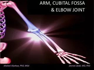
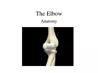
The humerus is a long bone in the arm that runs from the shoulder to the elbow.
It connects the scapula and the radius Body of humerus - cylindrical in its upper portion, and more prismatic below.
Lower extremity of humerus - consisting of a faceted condyle that articulates with the radius.
Upper extremity of humerus - consisting of a rounded head, a narrow neck, and two short processes, or "tuberosities". Distally, the capitulum of the humerus articulates with the head of the radius, and the trochlea of the humerus articulates with the olecranon process of the ulna.
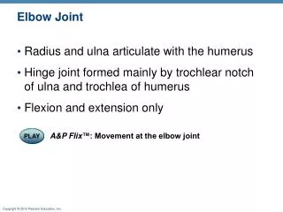
The grooved surface at the lower end articulating with the trochlear notch of the ulna. Above the back of the trochlea is a deep triangular depression, the olecranon fossa, in which the summit of the olecranon is received in extension of the forearm.
A bonyprominence of the outeraspect is the lateralepicondyle that providesorigin for the muscles which dorsiflex the wrist. Inflammation at this site is known as tennis elbow. medial epicondyle is larger and more prominent than the lateral epicondyle that gives attachment to the ulnar collateral ligament of the elbow-joint and ulnar nerve runs in a groove on the back of this epicondyle.
Above the front part of the trochlea is a small depression, the coronoid fossa, which receives the coronoid process of the ulna during flexion of the forearm.
The radius is the bone of the forearm that extends from the lateral side of the elbow to the thumb side of the wrist. The radius is situated on the lateral side of the ulna, It is a long bone, prism-shaped and slightly curved. The radius articulates with the capitulum of the humerus. Radius connects the elbow to the forearm.
Head of Radius, disk shaped prominence at proximal end of bone, forms articulating surface with capitulum of humerus. Radial tuberosity is a round projection that serves as an attachment point for Biceps Brachii muscle. Ulnar notch of Radius, slide depression that articulates with Ulna. Styloid process of Radius is the lateral projection at distal end of bone that forms lateral portion of wrist joint.
Ligaments of the elbow • Radial anular ligament • Interosseousmemebrane • Ulnar collateral ligament • Radial collateral ligament
Ligaments of the hand • Palmaraponeurosis • Flexor retinaculum
Bursae • Subcutaneous olecranon bursa • Subtendinousolecranon bursa
Cartilage • Articular cartilage
Articular Capsule • Synovial membrane • Fibrous layer (fibrous capsule)
Biceps • O: Scapula: Long head, supraglenoid tubricle, Short head, coracoid process • I: Radial tuberosity of radius • Brachialis • O: Distal half of humerus, anterior surface • I: Corocoid process and ulnar tuberosity • Pronator Teres • O: Medial epicondyle of humerus • I: Lateral aspect of radius at its midpoint • Brachioradialis • O: Lateral supracondylar ridge of humerus • I: Styloid process of radius • Supinator • O: Lateral epicondyle of humerus and adjacent ulna • I: Anterior surface of the proximal radius
Palmaris Longus • O: Medial epicondyle of humerus • I: Palmar fascia • Flexor Carpi Radialis • O: Medial epicondyle of humerus • I: Base of second and third metacarpals • Flexor Carpi Unlaris • O: Medial epicondyle of humerus • I: Pisiform, and the base of 5th metacarpal • Flexor Digitorum porfundus • O: Upper three-fourths of ulna • I: Distal phalanx of the four fingers • Pronator Teres • O: Medial epicondyle of humerus and coranoid process of ulna • I: Lateral aspect of radius at its midpoint
Flexor digitorum superficialis • O: Common flexor tendon, coronoid process and radius • I: Sides of the middle phalanx of the 4 fingers • Flexor Pollicis longus • O: Radius, anterior surface • I: Distal phanlanx of pollex • Abductor pollicis longus • O: Posterior radius, interosseous membrane, middle ulna • I: Base of the first metacarpal • Pronator Quadratus • O: Distal fourth of ulna • I: Distal fourth of radius
Extenson carpi radialis longus • O: Supracondylar ridge of humerus • I: Base of second metacarpal • Extensor carpi Ulnaris • O: Lateral epicondyle of humerus • I: Medial side of base of 5th metacarpal • Extensor Digitorum • O: Lateral epicondyle of humerus • I: Base of distal phanlanx of the 2nd-5th fingers • Extensor carpi radialis brevis • O: Lateral epicondyle of humerus • I: Base of 3rd metacarpal • Extensor digiti minimi • O: Lateral epicondyle of humerous • I: Base of distal phalanx of 5th finger • Extensor pollicis brevis • O: Posterior Distal radius • I: Base of proximal phalanx of pollex • Extensor pollicis longus • O: Middle posterior ulna and interosseous membrane • I: Base of distal phalanx of pollex
Triceps • O: Long Head: infraglenoid tubrecle of scapula, Lateral Head: Inferior to greater tubericle on posterior humerus, Medial Head: posterior surface of humerus • I: Olecranon process of ulna
Biceps: A: Elbow Flexion, forearm supination I: Musculocaneous nerve V:Brachial artery • Triceps: A: Elbow extention I: radial nerve, V:Deep brachial artery • Brachialis A: Elbow flexion I: Musculocutaneous nerve, V:brachialAtery • Brachioradialis: A: Elbow flexion I: Radial nerve, V:radial artery • Supinator: A: Forearm supination I: radial nerve, recurrent interosseous
Pronatorteres: A: forearm pronation, asstive in elbow flexion I:median nerve V:ulnar artery • Pronatorquadratus: A: forearm pronation I: median nerve V: anterior interosseous artery • Flexor carpiradialis: A: wrist flexion, radial deviation I: median nerve V: radial and ulnar arteries • Flexor carpiulnaris: A: wrist flexion, ulnardeviation I: ulnar nerve V: ulnar artery • Extensor carpiradialislongus: A: wrist extension, radial deviation I: radial nerve V: radial artery
Extensor carpiraialisbrevis: A: wrist extension I: radial nerve V: radial artery • Extensor digitorum: A: extends all three joints of the fingers I:radial nerve V: recurrent interosseous artery • Extensor carpiulnaris: a: extens and adducts wrist I: deep radial nerve V: ulnar artery • Flexor digitorumsuperficialis: A: Flexes MP and PIP jpints of the fingers I: Medial Nerve V: Ulnar artery • Flexor digitorumprofundus: A: flexes all three joints of the fingers I: Median and ulnar nerve A: Ulnaratrety
Flexor pollicislongus A: flexes all joints of the thumb I: Radial nerve V: posteriorinterosseous artery • Abductor pollicislongus: A: abducts pollucs I: radial nerve V: posterior interosseous artery • Extensor digitiminimi :A: extends all joints of the fifth finger I: Radial nerve V: Recurrent interosseous artery • Extensor pollicisbrevis: A: Extends MP joint of thumb I: radial nerve V: Posterior interosseousartery • Extensor Pollicislongus: A: extends the Mp and IP joints of the thumb I: Radial nerve V: Posrteriorinterosseous artery • Palmaris longus : A: Assistive in wrist flection I: Median nerve V: Ulnar artery