Download

1 / 22
230 likes | 563 Vues
Hypersensitivity reactions. Overview. Hypersensitivity, allergic reaction similar to protective mechanisms exaggerated and damaging to host Antigens = allergens Classified into 4 types (Gell & Coombs) Type I : Anaphlylactic reaction Type II : Cytolytic, cytotoxic reaction

E N D
Overview • Hypersensitivity, allergic reaction • similar to protective mechanisms • exaggerated and damaging to host • Antigens = allergens • Classified into 4 types (Gell & Coombs) • Type I : Anaphlylactic reaction • Type II : Cytolytic, cytotoxic reaction • Type III : Immune complex reaction • Type IV : Cell-mediated immunity (CMI), Delayed type hypersensitivity (DTH)
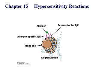
Hypersensitivity reactions: Antibody-mediated (type 1) reactions
Anaphylactic reaction • Anaphylaxis (Portier & Richer) • Glycerin extract of sea anemone induced exaggerated response on 2nd injection • ‘ana’ = away from, ‘pro’ = toward, ‘phylaxis’ = protection • Mediated by IgE Ab - bind through Fc portion with Fce receptors on mast cells & basophils • Three phases • Sensitization : IgE production upon Ag stimulation & binding of Fc on mast cells & basophils • Activation : Re-exposure to Ag & granule release • Effector : Anaphylaxis due to pharmacologic activity of released agents
Figure 14.1Electron micrograph of a normal mast cell illustrating the large monocyte- like nucleus and the electron-dense granules. On the right, a mast cell has been triggered and is beginning to release the contents of its granules, as seen by their decrease in opacity and the formation of vacuoles connecting with the exterior. [Photographs courtesy of Dr. T. Theoharides, Tufts Medical School.]
Sensitization • Exposure to allergens by mucosal contact, ingestion or parental injection resulting in IgE production • 50% of population produce IgE to air-borne allergens, only 10% develop clinical symptoms • ‘atopy’ (uncommon) = unique, unexpected response • atopic = affected patients • IgE production = T-dep. (TH2 cells - IL4) • Low level of IgE in non-allergic individuals = Ts & TNF-a
Skin testing by intradermal injection of allergens into the forearm
Activation • Triggering of mast cells to release granules & pharmacologically active components • Requires bridging of at least 2 receptors of IgE Tc
Figure 14.2Mast cell degranulation mediated by antigen-crosslinking of IgE bound to IgE Fc receptors (FceRI).
Figure 14.3Alternate ways in which mast cells can be induced to undergo degranulation.
Figure 14.4Mediators released during activation of mast cells.
Effector Phase • Principal clinical features • Swelling lips, tongue & larynx blocks respiration • Broncho-constriction prevent expiration • Dilation of blood vessels causes drop of blood pressure • Contraction of intestinal smooth muscles result in cramps & diarrhea • Increased vascular permeability causes urticaria
Figure 14.5A diagrammatic representation of late-phase reaction of type I IgE- mediated hypersensitivity with some of the mediators involved.
Figure 14.6Overview of induction and effector mechanisms in type I hypersensitivity.
Figure 14.7A diagrammatic representation of the destruction of a worm by eosinophils that have migrated to the area and been activated following IgE- and antigen- mediated mast cell degranulation.
Figure 14.8Electron micrograph (X6,000) of eosinophils (E) adhering to an antibody- coated schistosomulum (S). The cell on the left has not yet degranulated, but the one on the right has discharged electron-dense material (arrows), which can be seen between the cell and the worm. [Photograph courtesy of Dr. J. Caulfield, Harvard Medical School.]
Immunologic intervention • Hyposensitization • attempts to ‘desensitize’ with repeated low doses of allergens • Mode unclear : • Induction of ‘blocking’ Ab (IgG, IgA?) • Induction of tolerance due to switch from TH2 to TH1 or induction of Ts • Clinically altered allergens (allergoids)