HYPERSENSITIVITY REACTIONS
HYPERSENSITIVITY REACTIONS. HYPERSENSITIVITY REACTIONS. Innocous materials can cause hypersensitivity in certain individuals. leading to unwanted inflammation damaged cells and tissues. Non-proper reaction of the immune system to foreign substances


HYPERSENSITIVITY REACTIONS
E N D
Presentation Transcript
HYPERSENSITIVITY REACTIONS Innocous materials can cause hypersensitivity in certain individuals leading to unwanted inflammation damaged cells and tissues Non-proper reaction of the immune system to foreign substances Mainly harmless substances – after second or multiple exposure
TYPES OF ANTIBODY MEDIATED HYPERSENSITIVITY REACTIONS FcRIα)
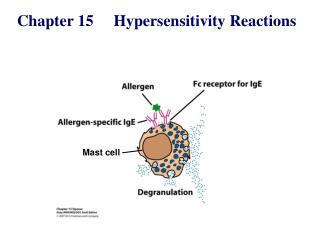
TYPE I HYPERSENSITIVITY REACTION ALLERGY
MAST CELL RESPONSE TO SURFACE FcRεI CROSSLINKING EARLY MEDIATORS Biogenic amins – histamin Enzymes – triptase, chymase, carboxypeptidase LATE MEDIATORS
THE EFFECT OF MAST CELL DEGRANULATION VARIES WITH THE TISSUE EXPOSED TO ALLERGEN
SYSTEMIC ANAPHYLAXIS IS CAUSED BY ALLERGENS THAT REACH THE BLOOD STREAM
Short/Common ragweed (Ambrosia artemisiifolia) Mugwort (Artemisia vulgaris) Green leaf back White leaf back
Mugwort (Artemisia vulgaris) – ? Wormwood (Artemisia absinthium) – Absinthe (thujone: max 35 mg/l)
MAST CELL DEGRANULATION, ALLERGIC REACTION IN THE SKIN OF A SENSIBILIZED INDIVIDUAL PRICK TEST
ImmunoCAP Specific IgE Blood Test Anti-IgE Serum IgE Allergen Solid phase
COMPARISON OF SKIN TEST TO SPECIFIC IgE TESTING Skin Pricktest Specific IgE allergens many almost all speed 20 min 1-2 days (result) medication no antihistamines no problem disease severe eczema: no problem difficult cost € 20 (total) € 20 per specific IgE sensitivity high slightly lower
Type II hypersensitivity IgG type antibodies bound to cell surface or tissue antigens • cells expressing the antigen become sensitive to complement mediated lysis or to opsonized phagocytosis • frustrated phagocytosis tissue damage • the antibody inhibits or stimulates target cell function no tissue damage (e.g. M. gravis – receptor-blocking antibodies)
FRUSTRATED PHAGOCYTOSIS MEDIATED BY IgG TYPE ANTIBODIES Binding Opsonization Internalization Enzyme release The tissue, which can not be phagocytosed, is damaged Absorbed antigen (drug) Opsonized surface Binding Frustrated Enzyme release phagocytosis
EXAMPLES - TYPE II HYPERSENSITIVITY Newborn haemolytic anaemia Transfusion reaction Hyperacut allograft rejection Drug-derived • Haemolitic anaemia • Thrombocytopenia • Agranulocytosis • Penicillin-based antibiotics • Anti-arithmic quinidine Goodpasture syndrome (kidney, basal membrane - collagen type IV) Pemphigus vulgaris (mucosal bubbles) against desmosomal antigens, interruption of epidermal and mucosal connections, acantolysis (desintegration into single cells) Myasthaenia gravis (anti-acetyl-choline receptor antibodies) Graves-Basedow-Flajani disease (anti-TSH-receptor antibodies)
TYPE III HYPERSENSITIVITY Antibodiesbinding to solubleantigens forming small circulating immune complexes which are deposited in various tissues Depends on: Size of immune complexes Antigen-antibody ratio Affinity of antibody Isotype of antibody
THE PROCESS OF TISSUE DAMAGE CAUSED BY IMMUNE COMPLEXES Blood vessel wall permeability Frustrated phagocytosis Immune complexes activate the complement system, neutrophils, basophils and thrombocytes
SYPMPTPOMES CAUSED BY TYPE III HYPERSENSITIVITY REACTIONS DEPEND ON THE SITE OF IMMUNECOMPLEX DEPOSITION
ARTHUS-REACTION • Localized Type III hypersensitivity • Local vasculitis develops as a result of immune complex deposition • Inhaled antigens (fungi, animal feces) may induce similar reaction in the lung • IgG type antibody • ‘Farmers lung’ and ‘piegeon-breeder’s lung’
MANIFESTATION OF TYPE III HYPERSENSITIVITY IN LUPUS ERYTHEMATOSUS Facial, malar "butterfly" rash with characteristic shape across the cheeks. Discoid lupus erythematosus (DLE) involves mainly the skin, it is relatively benign compared to systemic lupus erythematosus (SLE). In either case, sunlight exposure accentuates this erythematous rash. A small number (5 to 10%) of DLE patients go on to develop SLE (usually the DLE patients with a positive ANA). Here is a more severe inflammatory skin infiltrate in the upper dermis of a patient with SLE in which the basal layer is undergoing vacuolization and dissolution, and there is purpura with RBC's in the upper dermis (which are the reason for the rash).
ANA Anti-nuclear antibody
TYPE IV HYPERSENSITIVITY REACTION T CELL MEDIATED PROCESS
DELAYED-TYPE HYPERSENSITIVITY(DTH) (e.g. tuberculin skin test) TH1 from a previous immunization (memory)
Tuberculin skin test Introduction of Ag Ag = antigen Purified protein derivate (PPD)
DTH as a result of a contact-sensitizing agent* CONTACT DERMATITIS *a contact-sensitizing agent is usually a small molecule that penetrates the skin then binds to self-proteins, making them “look” foreign
Poison ivy Anacardiaceae (family), Toxicodendron (genus) Toxicodendron radicans or Rhus toxicodendron