Download
1 / 53
700 likes | 1.63k Vues
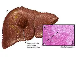
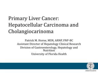
Primary Liver Cancer: Hepatocellular Carcinoma and Cholangiocarinoma. Patrick M. Horne, MSN, ARNP, FNP-BC Assistant Director of Hepatology Clinical Research Division of Gastroenterology, Hepatology and Nutrition University of Florida Health. Disclosures.

E N D
Primary Liver Cancer: Hepatocellular Carcinoma and Cholangiocarinoma Patrick M. Horne, MSN, ARNP, FNP-BC Assistant Director of Hepatology Clinical Research Division of Gastroenterology, Hepatology and Nutrition University of Florida Health
Disclosures • Financial relationships to disclose within the past 12 months: • Grant support with Bayer/Onyx
Objectives • Discuss the diagnosis and management of hepatocellular carcinoma (HCC) and cholangiocarinoma • Review current screening and staging of both HCC and cholangiocarinoma • Selection of the optimal, evidence-based surgical and nonsurgical treatment modalities • Review current treatment options
Background of HCC • World wide • 6th most common cancer – 748,271 new cases per year • 3rd leading cause of cancer-related mortality – 695,843 deaths per year • US incidence has tripled over the last three decades • Estimated new cases: >20,000 new cases annually • Fastest Growing Death Rate in the US • Dismal 5-year survival 10% • 80%-90% of HCC cases occur in cirrhotic livers • Leading cause of death in cirrhosis Globocan 2008. McGlynn KA et al. Int J Cancer. 2001;94:290-296; McGlynn KA et al. Cancer Epidemiol Biomarkers Prev. 2006;15:1198-1203; El-Serag HB. Gastroenterology. 2004;127:S27-S34; Altekruse SF et al. J ClinOncol. 2009;27:1485-1491
U.S.-HCC Risk Factors Patients with HCC Distribution of Markers N = 239 HCV HBV Alcohol Cryptogenic Other El-Serag HB, Rudolph KL. Gastroetnerology2007;132:2557-2576. SnowbergerN, et al. Alim Pharm Ther 2007;26:1187.
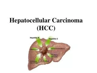
Risk Factors for HCC • Host Factors • Cirrhosis • HBV, HCV, NASH, Alcohol • Hemachromatosis, • α-1 antitrypsin deficiency • HBV • Male gender • Family history of HCC • Coinfectionwith HCV, HBV, or HIV • Older age • Viral Factors • HBV DNA levels >2,000 IU/mL(10,000 copies/mL, REVEAL Study) • HBV genotype C* • Basal core promoter mutation • Presence of HBeAg • History of reversions fromanti-HBe to HBeAg • Likely Factors • Diabetes • Obesity AASLD practice guidelines. AASLD website. Available at: http://www.aasld.org/practiceguidelines. Accessed 02/10/11; Keeffe. ClinGastroenterolHepatol. 2008;6:1315; Mahtab. HepatobiliaryPancreat Dis Int. 2008;7:457; Lin. Hepatology. 2007;46:1034.
HCC Pathogenesis Farazi PA and DePinho RA. Nat Rev cancer 2006.
HCC Incidence has TripledDismal 5 year survival (~10%) SEER = Surveillance, Epidemiology, and End Results. AltekruseSF et al. J ClinOncol. 2009;27:1485-1491
Groups for Whom HCC Surveillance is Recommended AASLD Practice Guidelines • Cirrhosis • Viral hepatitis – HCV, HBV • Non-alcoholic fatty liver/non-alcoholic steatohepatitis (NAFLD/NASH) • Alcohol and other • Hepatitis B carriers • Asian males over 40 yrs • Asian females over 50 yrs • All cirrhotic hepatitis B carriers • Positive family history of HC • Non-cirrhotic HBV carriers with high HBV DNA levels or more severe current/past levels of inflammatory activity AASLD=American Association for the Study of Liver Diseases; HBV=hepatitis B virus. Practice guidelines page. AASLD website. Available at: http://www.aasld.org/practiceguidelines.
Current Recommendation for Surveillance • AASLD practice guidelines • Ultrasound every 6 months • Goal • Detect early tumor and improve survival • AFP • Normal in 40% of patients with HCC • Inadequate sensitivity and specificity in the diagnostic range – normal in small HCC • Values >200 ng/mL high PPV in cirrhotic patients with a mass El Serag. Gastroenterology. 2008;134:1752; Practice guidelines page. AASLD website. Available at: http://www.aasld.org/practiceguidelines. Accessed 10/12/11; Sheu. Gastroenterology. 1985;89:259.
Case #1 • 67 year old white male with HBV cirrhosis on antiviral therapy with good viral control. • Abnormal US on surveillance. • AFP = 5. • Cirrhotic liver, stigmata of portal hypertension. Single 2.5 cm arterially enhancing lesion in the left lobe of the liver.
Diagnostic Algorithm for HCC in Cirrhosis New Liver Nodule on US <1 cm >1 cm Repeat US at 3 months 4-phase MDCT / dynamic contrast enhanced MRI Arterial hypervascularity AND venous or delayed phase washout Stable Growing/changing character Other contrast enhanced study (CT or MRI) No Yes Investigate according to size HCC Arterial hypervascularity AND venous or delayed phase washout Biopsy Adapted from Bruix J, Sherman M. Hepatology July, 2010. Available at http://www.aasld.org/practiceguidelines/Pages/NewUpdatedGuidelines.aspx. Accessed 08/03/10. No Yes
Radiologic Diagnosis of HCC in Cirrhosis Arterial phase enhancement Venous phase “washout” Cabrera R, Nelson DR. Aliment PharmacolTher. 2010 15;31(4):461-76.
Child-Pugh Scoring for Cirrhosis INR=international normalized ratio. Child, ed. The Liver and Portal Hypertension. Philadelphia, PA: Saunders;1964:50-64; Pugh. Br J Surg. 1973;60:646. D’Amico G et al. J Hepatology 2006; 44:217-231.
Model for End-Stage Liver Disease (MELD) Predicts Severity of Liver Disease • Predicts severity of liver disease and cirrhosis-related mortality (3 month survival) • Used in allocation of organs for transplant • Originally designed to predict survival after TIPS procedure • Mortality by MELD score • 10-19: 8% • 20-29: 24% • 30-39: 60% • >40: 81% Weisner RE et a. Gastro 2003. Kamath P and Kim WR. Hepatology. 2007.
New Classification of Cirrhosis D’Amico G J. Hepatology 2006;44:217-231; Arvaniti V et al Gastroenterology 2010;139:1246-1256.
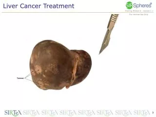
Surgery: Liver Transplantation Liver Resection Ablation: Percutaneous ethanol injection (PEI) Radiofrequency (RFA) Transarterial: Chemoembolization 90Y microspheres Systemic Therapies: Sorafenib Clinical Trials What is the best treatment option for this patient? CURATIVE Palliative
Liver Transplantation for HCC: Milan Criteria • 5-yr survival with transplantation: ~ 70% • 5-yr recurrent rates: < 15% Single tumor, <5 cm Up to 3 tumors, all <3 cm Absence of macroscopic vascular invasion and extra-hepatic spread Mazzaferro V, et al. N Engl J Med. 1996;334:693-699. Llovet JM. J GastroenterolHepatol. 2002;17(suppl 3):S428-S433.
Patient Survival of HCV LT recipientswith HCC and without HCC Cabrera R et al. American Journal of Clinical Oncology. 2012 Aug;35(4):345-50.
Candidates for RFA and PEI • Includes individuals who are not candidates for surgery – resection or transplantation • Radiofrequency ablation generally preferred over percutaneous ethanol injection • Necrotic effect more predictable across tumor sizes • Meta-analyses (4 RCTs) suggest better local control and survival benefit with RFA vs PEI Bruix J, et al. AASLD HCC Guidelines. July 2010.
New Trends in Local Ablation • Microwave ablation • Irreversible electroporation • Intravenous heat-sensitive liposomal doxorubicin in combination with RFA • Combination chemoembolization and RFA
Case #2 • 70 year old white male with HCV cirrhosis noted with abnormal ultrasound. • AFP =11. • Cirrhotic liver multiple arterially enhancing lesions in the right and left hepatic lobes. 2 large enhancing lesion in the superior right hepatic lobe which measure 5.6 x 4.9 cm and 5.7 x 5.6 cm.
Randomized Study of Conventional TACE vs DEB TACE Lammer J, et al. CardiovascInterventRadiol. 2010;33:41-52.
Contraindications to TACE • Extrahepatic tumor spread • Lack of portal blood flow • Portal vein thrombosis, portosystemic anastomoses or hepatofugal flow • Advanced liver disease (Child-Pugh Class B or C) • Clinical symptoms of end-stage cancer
Radioembolization Using Yttrium-90 Single institution cohort study • Endpoints: Response rate,* TTP,* survival, toxicity • 273/291 (94%) of patients had follow-up imaging • 58% Downstaged, 32 transplanted • Response rate 42% (WHO) and 57% (EASL) • No GI ulcers Salem R et al. Gastroenterology. 2010;138(1):52-64.
New Trends in Loco-Regional Therapy • Doxorubicin-eluting beads • Quadraspheres • Combination TACE or Y90 with sorafenib • SBRT and Proton beam therapy
Case #3 • 47 year old white male with HCV cirrhosis undergoes a screening abdominal ultrasound. • Ultrasound reveals multiple liver masses. • Innumerable bilobar arterial enhancing lesions and vascular invasion. AFP >50,000. consistent with diffuse HCC
UF Experience with Sorafenib in HCC (n=31) 1.0 0.8 0.6 Cum Survival 0.4 0.2 0.0 0 4 8 12 16 20 24 28 32 36 40 44 48 Survival time in weeks Overall Survival Curve - Median Survival of 36 wks (9 months) Cabrera R, et al. 2008 Gastrointestinal Cancers Symposium. Abstract 147.
UF Experience: Sorafenib in HCC Sorafenib 400 mg PO BID N = 31 Child-Pugh Class A = 64%, B = 36% BCLC Stage B 23%, Stage C 77% Cabrera R, et al. 2008 Gastrointestinal Cancers Symposium. Abstract 147.
GIDEON Observational HCC Study • Differences in usage and reported toxicities • Patients with Child-Pugh B experienced more liver-associated AEs • Median TTP • Child-Pugh A, 4.2 months • Child-Pugh B, 3.6 months • Median OS: • Child-Pugh A, 10.3 months • Child-Pugh B, 4.9 months OS= overall survival; TTP=time to progression. LencioniInt J ClinPract 2012; Venook. ASCO GI. 2011 (abstr 157); Marrero ASCO. 2011 (abstr 4001).
TACE in Combination With Sorafenib Sorafenib 400 mg PO BID TACE N = 48 Child-Pugh Class A = 72%; Child-Pugh Class B = 28% BCLC Stage B 81%; Stage C 19%. Overall Median Survival 18.5 months (Child B 17.6 months; Stage C 17.0 months) Cabrera R et al. Aliment PharmacolTher. 2011;34(2):205-13.
Investigational Treatment Strategies Cabrera R. Clinical Liver Disease. Dec 2012.
Cholangiocarcinoma (CCA) • Background: • Arises from the epithelial cells of the bile ducts. • Rare in the United States.
Worldwide prevalence Bridgewater J J. of Hepatology2014
Risk Factors for Cholangiocarcinoma • Primary sclerosing cholangitis (PSC) • Almost 30% of cholangiocarcinoma case are in the setting of PSC. • Annual incidence is between 0.6-1.5%. • Lifetime risk is 10-15%. • Fibropolycystic liver disease • Parasitic infection • Viral hepatitis • Though much lower than risk for HCC Burka K. Am J Gastroenoterology 2004
Incidence of Cholangiocarcinoma Cumulative incidence of Years since PSC diagnosis Courtesy of Dr. Keith Linder, MD., ASU
AASLD guidelines • Risk factors for developing CCA • Elevated serum bilirubin • Variceal bleeding • Proctocolectomy • Chronic UC with colorectal cancer or dysplasia. Practice guidelines page. AASLD website. Available at: http://www.aasld.org/practiceguidelines.
Biliary strictures • Signs and symptoms: • Jaundice, pruritis, abdominal pain, fever, weight loss • Cholangitis is unusual. • Tumor markers? • CEA • CA 19-9
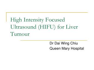
Imaging diagnosis of CCA • Challenging to say the least • Ultrasound • CT scan • MRI with MRCP • ERCP with brushing • No clear guidelines as to best modality to use or frequency
Practice guidelines page. AASLD website. Available at: http://www.aasld.org/practiceguidelines.
Case 1 • 50 year old male with known history of PSC undergoes routine ultrasound screening. Follow up MRCP
Treatment options • Criteria for surgical resection: • Absence of retropancreatic and paraceliac nodal metatases or distant liver metastases • Absence of invasion of portal vein or main hepatic artery • Absence of extrahepatic adjacent organ invasion • Absence of disseminated disease • Ultimately, candidacy for resection is determined at surgery! Tsao JI et al Ann Surg. 2000 Su CH et al Ann. Surg. 1996
Distal Extrahepaticcholangiocarcinoma • Considered having the highest rate of resection. • Role of adjunct therapy post resection, commonly recommended but lack of data determining if true benefit.
Case 2 • 60 year old female presents to the local Emergency Department with sudden onset painless jaundice
Role of Radiotherapy with or without chemoradiotherapy • Approaches include external beam irradiation (ERBT) alone or… • In combination with chemoradiotherapy • Most commonly used: • 5-FU • Gemcitabine • Cisplatin • oxaliplatin