Download
1 / 70
720 likes | 935 Vues
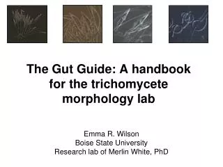
The Gut Guide: A handbook for the trichomycete morphology lab. Emma R. Wilson Boise State University Research lab of Merlin White, PhD. Introduction Part 1: Overview of Trichomycetes Symbiosis Taxonomy Part 2: Collecting Specimens Where are they? Aquatic collections Sorting Hosts

E N D
The Gut Guide: A handbook for the trichomycete morphology lab Emma R. Wilson Boise State University Research lab of Merlin White, PhD
Introduction Part 1: Overview of Trichomycetes Symbiosis Taxonomy Part 2: Collecting Specimens Where are they? Aquatic collections Sorting Hosts Part 3: Maintenance, Dissection & Preservation Keeping hosts alive Dissection Host preservation Fungal preservation Part 4: Culturing Media recipes Making media Pouring Plates Making Slants Transferring cultures Part 5: Morphometrics Where and what to measure Examples and tips Conclusions Appendices 1. Species list by Order 2. Host keys 3. Further reading Contents
Introduction • This handbook is made for any student who would like to deepen their understanding of the curious world of trichomycete fungi and protists*. • We will start out by briefly introducing the biology of trichomycetes. • Next, we’ll give some examples and tips for specimen collection and how to be successful in the field. • After that we’ll focus on the tricho hosts and what to do with them once you’ve gathered them in the field. • One important aspect of trichomycetology is culturing. That will be covered in Part 4 of the guide. • Finally, we’ll go over morphometrics (a way to measure your tricho and determine what species you have). • At the end of this guide, you’ll find several appendices including a list of genera, host keys, and a link for further reading. Enjoy! * “trichomycete fungi and protists” will often be shortened to “trichos” (pr: “trick-Ohs”) throughout this handbook.
Web Resources • In addition to this guide, the following links are vital for anyone interested in trichos: • http://www.nhm.ku.edu/fungi/ • http://www.nhm.ku.edu/fungi/Lucid%20Keys.html • http://www.nhm.ku.edu/~fungi/Monograph/Text/Mono.htm • Created by Dr. Robert Lichtwardt – the modern father of trichos. • Lets go through each section to show some highlights of how best to use each page as a burgeoning trichomycetologist.
Shortcut to Lucid Keys Shortcut to Monograph Web Resources • On the home screen of http://www.nhm.ku.edu/fungi/ , there is a brief summary about trichos as well as two very useful links – the Monograph and the Lucid Keys – that are used by scientists in the lab nearly every day. • We’ll go into detail about these next
Contents are a great place to start navigating Glossary contains many tricho-specific definitions Monograph • This page is essentially a book (originally published in 1986) whose current version is published on the web. • It contains a wealth of information from host specificity to molecular techniques with trichos • The easiest way to navigate the monograph is to click on “Contents” and choose from the listing on the page that appears. • There is also a great glossary for many tricho-specific terms.
A B Lucid Keys • Once you have a trichomycete in front of you, how do you know what it is? • The best place to quickly identify your tricho is to navigate to the Lucid Keys • You will see a list (A) to choose from • If you have a fungal species, the choices highlighted in pink are the ones to examine. If you’re dealing with a protist, choose one of the other links. • Once you’ve clicked on your link, a screen appears (B) where you can input measurements, hosts, gut attachment, etc. and retreive a list of species that match your features. • This will all become more clear as you go through the handbook and familiarize yourself with trichos. We will revisit this sight in Part 5 – Morphometrics.
Part 1 Overview of Trichomycetes Harpellales Asellariales Amoebidiales Eccrinales
Overview* • Trichomycetes are obligate endosymbionts of a variety of arthropods. • Their hosts may be immature aquatic stages of insects, as well as adult terrestrial arthropods. • Trichos reside in the digestive tract of these organisms commensally – that is, they have a neutral relationship with their host. However, there have been several studies showing that they can shift between all aspects of symbiosis including mutualism and parasitism. • There are currently 385 species of trichos (including both fungal and non-fungal members). • Their systematics is in a state of flux, but molecular phylogeneticists are in the process of stabilizing this group of organisms in order to strengthen the base of the fungal tree of life. *This section is decidedly brief. Refer to the monograph and further reading (Appendix III) for more information.
Shifting continuum Overview - Symbiosis • Symbiosis refers to two or more organisms living in close association with each other. It spans from parasitism through commensalism to mutualism. • Interestingly, trichomycetes exist along the entire spectrum of symbiosis. Symbiosis Parasitism Commensalism Mutualism Host Symbiont – + Host Symbiont ø ø + + Host Symbiont Effects Tricho example Smittium morbosum§ Default for all other trichos Smittium culisetae ¥ ¥Horn & Lichtwardt, 1981, Mycologia 73: 724-740 §Sweeney, 1981, Transactions of the British Mycol Soc, 77: 55-60
Overview – Taxonomy • The Class Trichomycetes were described by R.W. Lichtwardt in 1954. • All members were originally described as fungi (trichomycete translates to “hair fungus” because of the hair-like nature of the organisms in the gut) • Traditionally there were four orders described: Amoebidiales, Asellariales, Eccrinales and Harpellales. • In 2002 it was confirmed by Mendoza¥ that the Amoebidiales were non-fungal, and are now considered ichthyosporean protists. • In 2005, using molecular phylogenetics, Cafaro§ found that the Eccrinales were also protists. • The Harpellales and Asellariales are true fungi, yet their phylogenetic placement as early diverging members of the fungal tree of life is in flux. • Currently, scientists are resolving the issues of many clades and finding new relationships among taxa using new molecular technology. • Later in this guide you will learn how to preserve a fungal specimen so that its DNA can be extracted for analysis. ¥Mendoza, Taylor & Ajello, 2002, Ann Rev Microbiol, 56:315-344 §Cafaro, 2005, Molecular Phyl & Evoln, 35: 21-34
Taxonomy Here is a table of some non-genetic features that separate the fungi from the protists
Part 2 Collecting Trichomycetes Pictures are from sampling at Dry Creek, February, 2011. From left to right: PrasannaKandel, Emma Wilson, Nicole Reynolds, and Zach Hoefer.
Collecting Trichomycetes • It is a relatively simple process to collect trichomycete hosts from nature • Although there are many hosts of trichos, this section will focus on methods for collecting one of the true fungal members of the group: Harpellales* • We’ll provide locations that hosts are likely to be found, a list of supplies needed for aquatic sampling, and methods on how to sort the “good” from the “bad” aquatic insects. *Whenever you collect Harpellales, non-fungal members in the genus Paramoebidium are often found in the same hindgut
A C Where are trichos found? • Where aren’t they found?! • Most immature aquatic insect hosts require flowing water, so streams are a great place to find hosts. • Aquatic hosts are found everywhere from pristine streams (A) to irrigation ditches (B) • Often, shed skins (exuvae) of hosts contain trichomycetes, so collecting in pools where they’ve washed up may also prove fruitful (C). B Reynold’s Creek, Idaho Sarah Oman in Parma, Idaho Emma Wilson and Yan Wang, Boise River, Idaho
http://www.bioquip.com/prod_images/7512D-001-Two-Piece-HD-Aquatic-Net.jpghttp://www.bioquip.com/prod_images/7512D-001-Two-Piece-HD-Aquatic-Net.jpg http://www.escalemodels.com/forums/uploads/monthly_08_2011/post-2198-094235900%201312467236.jpg http://www.babiestravellite.com/mm5/graphics/subcat_images_sq/Ziploc-Bags.jpg http://img.alibaba.com/img/pb/477/433/362/362433477_209.jpg http://www.tritechresearch.com/items/T3361-L.JPG http://image.become.com/imageserver/s1/901007951-75-75-5-32/stream-thermometer.jpg http://c1903.r3.cf3.rackcdn.com/YI132O770_1.jpg Collecting Freshwater Aquatic Hosts Prasanna Kandel showing off his hip waders! • For sampling aquatic hosts you’ll need the following items: • Boots (hip or chest waders work well) • Life vests for deep water • Nets (D-nets usually work best) • Buckets (optional) • Resealable plastic bags and/or tupperware containers • Transfer pipettes and forceps • 100mm Petri dishes (for black fly larvae) • Plastic pans • Stream thermometer • Cooler with ice for transport to the lab
Collecting Freshwater Aquatic Hosts • When sampling from lotic habitats (flowing water), its best to stand upstream from your net and use one of your feet to overturn rocks, kick leaves, and disrupt the area to bring insects up from the bottom. • Then, swish your net in a figure eight to capture all of the debris that is floating downstream (including insects!) • Put a bit of water in your pan, then shake your net into it. • You can then sort the desired insects from the large pan into your small tupperware containers, or simply pour your pan into the plastic bags and sort in the lab. • Put your tupperware or plastic bags on ice and head back to the lab. Yan Wang collecting hosts in the Boise River Emma Wilson sorting insects with students at Reynolds Crk Prasanna Kandel and Lance Steele sorting insects at Cottonwood Crk Sarah Oman and Emma Wilson packing samples into the cooler in Parma, ID
Collecting Black flies (Simuliidae) • Black flies are lower dipterans that are a great host of trichos (see guide in Appendix II). • Depending on stream conditions, you may not capture all of the available black flies because they tend to cling to substrates such as dangling vegetation • Therefore, it helps to swish the net through or pull up manually any trailing vegetation. • They may also be attached to rocks and boulders, so bringing them out of the stream and hand picking is also useful. Mass of black fly larvae on a twig from Dry Creek, Idaho
The Bad The Good http://academics.smcvt.edu/Vermont_rivers/River%20sites/Cold%20Brook.htm Sorting Aquatic Hosts • So now you have your insects in the pan – which ones do you take back to the lab? • In Appendex II you can find keys to identifying hosts to the family level. • The “bad” stonefly pictured above is in the family Perlidae, which are predaceous. • Not only are trichos not found in predaceous insects, but the good hosts (like the Nemouridae above) could be eaten by the undesirable hosts. Hmm... Which ones to take? Dr. White ponders which insects to collect … and perhaps the meaning of life?
Sorting Aquatic Hosts • Sorting hosts may be done in the field or in the lab. • The keys in Appendix II will help to identify the correct (i.e. non-predaceous hosts) • Using your pipette, transfer insects from the large collecting pan to the smaller tupperware pans, partially filled with stream water. • If the host is too big for the pipette, use the feather-tipped forceps to move them. • Black flies have very high oxygen demands, so if they are to be placed in deep water, they don’t survive as well (unless the pan is aerated upon return to the lab). • Therefore, it helps to place black fly larvae in petri dishes with a very thin film of water over them (see bottom picture). • This way, they can still remain moist, and have enough gas exchange through the small amount of water. From left to right: Mayfly, stonefly, black fly, midge http://www.lifeinfreshwater.org.uk/Species%20Pages/Midge_Chironomus.jpg.html Emma Wilson sorting hosts with high school kids at Reynold's Creek, Idaho
Part 3 Maintenance, Dissection and Preservation
Maintenance, Dissection and Preservation • A good deal of work in the morphology lab deals with maintaining, dissecting, and preserving hosts and trichos. • This section will guide you through this process in the following sections: • Documentation of samples • How to keep hosts alive in the lab • Dissection example and explanation • Preservation of host • Preservation of trichos (slides and CTAB)
State, Province or Country code (usually 2-3 letters) Serially increasing site reference number. May be accompanied by a letter to designate sub-sites in the location (e.g. “ID-55A”) Documentation of Samples • For each collection event, a unique collection code should be made as follows, separated by a hyphen: ID-55 • The sample code should be recorded in your field and lab books, or collection log, along with any relevant site information (stream temp, vegetation, substrate, GPS).
Maintaining Hosts in the Lab • The last thing you want, is for your sample to quickly die after returning to the lab. • The best way to prevent this is to dissect your hosts as quickly as possible. • Another approach, detailed here, is to create livable conditions for them in the lab itself. • It helps to keep black flies in a petri dish with a thin water overlay for short term storage, as previously described. An advanced setup with manifolds and airlines pumped through air diffusers to maintain hosts in the lab.
Maintaining Hosts in the Lab • For long term storage of hosts, a simple aeration system can be implemented. • You will need: • Air-diffusing stones • Air tubing • Air pump • Tupperware containers • Cut a small hole in the top of the tupperware and run a hose through, connected to an air pump. • Keep the sample in the fridge with the air pumping through the stone. • If the food looks depauperate, they can be fed a fish-food slurry (ground up tetra-fin in water). Tupperware container Air pump Tubing with airstones attached
Just before dissection… • They’re often really small! • Keep your hosts on ice in either a tupperware container or petri dish. • This will keep them cool and calm.
Dissecting Some useful supplies • For the most complete dissecting experience, you will need the following: • Stereo-microscope (one with dark-field lighting is best) • Glass slides (3”x1”x1.0mm) • Coverslips (glass 18x18 mm is best) • Fine tipped jeweler’s forceps • Insect mounting needles held by pin vices • Droppers of • Distilled Water • Lactophenol Cotton Blue (LCB) • NanoPure water • Small vials for host specimens • Also, small pieces of paper for vials • CTAB [Cetrimonium bromide ((C16H33)N(CH3)3Br] – a DNA buffer • 70% Ethanol (EtOH) • Delicate task wipers • A lab notebook • Pen and pencil i j a h b f k g c l d e
Dissecting • To begin with, take a clean glass slide and place two drops of water on it • The one on the left will be your “kill drop” • This is where you’ll be dissecting the gut out of the host • The one on the right will be your “dissection drop” • This drop may be preserved for future identification • Therefore, don’t put the drop too far to the right, otherwise it may not image very well • Place your host in the “kill drop” and begin dissecting. “kill drop” “dissection drop”
Dissecting – Dipterans Step 1 – Remove head and anus We’ll be using a black fly to demonstrate how to dissect a dipteran host. A A B C • Using one forcep, grab some exoskeleton on the section just below the head capsule – try to pin the larva down. With the other forcep, pinch the area to cleanly remove the head. • The anus can be seen by an “X” on the dorsal side of the larva. Its usually best to penetrate the insect with forceps and tear the anus open. • Look for the peritrophic matrix (clear tube coming from the body cavity – yellow star above, white arrow in facing picture). Often, the hindgut will protrude from the anal opening (black star above). B C
foregut Peritrophic matrix with food inside The intact midgut Lifting PM to remove food, using needle Midgut fungi observed as tiny hyaline hairs Dissecting – Dipterans: Midgut 1. 2. D E F G 3. • D. Once the gut is exposed, you can begin to remove the midgut, or peritrophic matrix (PM) • The peritrophic matrix is a clear “tube” that lines the inside of the midgut. • At the top, you can often see the foregut (image 1). • No gut fungi are found in the foreguts of dipterans, so it is best to remove the clear lining, which is often filled with food (image 2). • To remove the food, slide the needle underneath the PM, lift and lower it until it is as empty as you can get it (image 3). You may need to add more water to your slide to get all of the debris out. • Once the food is out, place the PM in a new drop of water (usually on a new slide) to image the gut fungi. Under the dissecting microscope, the trichos appear as small clear hairs (image 4). 4.
Dissecting – Dipterans: Hindgut 1. D The intact hindgut E F G 2. • F. To isolate the hindgut, detach the gut just posterior to the malpighian tubules, as pictured above with the black arrow (F). • The intact hindgut (image 1) is a coiled gut that is slightly opaque. • - Place the hindgut in the dissection drop, and use the needles to tear the gut open and into smaller pieces to release all of the trichos - both fungal and protistansymbionts may be present (image 2). • The dissection drop is often full of food, debris, and “host bits”. The example specimen had a Paramoebidium (non-fungal associate) in the hindgut - pictured in the circle (image 3). • If a tricho is found, you may mount it on a separate slide by picking up the fungus with the needle (similar to how the PM was picked up on the previous page) and placing it in a new drop. Using needles to tear open the hindgut and expose fungi 3. Post-dissection. Can you see the tricho in the circle?
Dissecting – Non-Dipterans We’ll be using a mayfly (Ephemeroptera, Baetidae) to demonstrate how to dissect a non-dipteran host. A A B C B D C The intact hindgut D Non- dipteran hosts (such as stoneflies and mayflies) do not require dissection of the midgut. They are dissected in a similar way to dipterans as far as head removal (A), but the hindgut may be dissected out by grasping the abdominal segment just anterior to the rectum and pulling (B), exposing the hindgut and malpigian tubules (arrow, C). The gut may then be dissected in the same fashion as described in the previous section (D) using the needles. One thing to be careful of is the extrusion of some species of trichos outside the anus (yellow star, C). When this happens, take extra care to not disrupt them as they often have produced sexual spores. You can place the entire anal segment on the slide, or try to remove them with your forceps and make a new slide.
Slide Preparation ID-55 State, Province or Country code (usually 2-3 letters) Serially increasing site reference number. May be accompanied by a letter to designate sub-sites in the location (e.g. “ID-55A”) • Now that you have your tricho on a slide, what do you do with it? • Carefully place the coverslip on the slide, avoiding air-bubbles and having the specimen squish out from underneath. If your slide is really wet, wait a few minutes for it to dry before proceeding. • Using a slide-specific marker, write the collection code followed by your initials and the number you’re on. • For instance, in the slide pictured, it was collection number 145 in Idaho (ID-145), and the dissector’s (Emma) first host (E1). • Also write: • Host ID (common name is fine) • Date • What is on the slide (midgut, hindgut, dissection drop) • The genus and species (if you know it)
Slide Preparation A • After writing the description on the left side of the coverslip, add a tiny drop of lactophenol cotton blue (LCB) to the right side of the coverslip. • As shown in the top panel of images (A), its best to completely empty the dropper of LCB to get a small droplet at the base. • This can then be applied to give just the right amount (B). • Next, apply a coat of clear nail polish (we use Revlon Clear, #771) over the text so that it doesn’t wash away while prepping the slide (C). • The LCB is then allowed to infiltrate the coverslip (D) for a few minutes to days. • The slide is kept flat (don’t ever tilt the slide until it is completely fixed!) on a letter tray while it is being prepared (E) . B C D E
Slide Preparation A B • The LCB is completely infiltrated when blue can be seen through the whole coverslip (A). • To wick off any excess LCB, a small piece of Kimwipe can be placed on the right side of the coverslip where the LCB drop was originally placed (B). • This can be left for a few hours to days. • Once the excess has been removed, clear nail polish is applied around three sides of the coverslip (C) • The right side is left bare (where the LCB was applied) (D) so that it may be rinsed in the next step. C Right side is left bare D
Slide Preparation • Once the nail polish has dried, the excess LCB needs to be rinsed off with distilled water. • Make sure that the slide is tilted to the right so the LCB rinses away from the text (see image). • After the slide dries, apply nail polish to all 4 sides of the coverslip. • Nail polish should be applied until there is a smooth transition between the slide and coverslip • You should be able to rub your finger along the nail polish and not be able to feel the sharp margin of the coverslip. • Once it is completely sealed, the slide can be put away in the appropriate slide box, and saved for future inspection. • Store the slide with the coverslip facing UP so you are working with gravity! • Remember, the more thorough the slide preparation, the longer the slide will last! Apply to all sides after rinsing
A Moist Chamber • Sometimes when you have a tricho, it helps to place the slide in a moist chamber to facilitate detachment of spores and the development of other features. • You will need the following items (A): • Filter paper – Petri dish • Distilled water – Bent glass rod • To make the moist chamber… • Place the filter paper in the petri dish (B) • Squirt a small amount of water into the dish (C) • Place glass rod on top of filter paper (D, E) • Put slide (with coverslip on and some extra water on the right side of coverslip) on top of the bent glass rod (F). • Cover petri dish, and label the isolate number, what is on the slide, date and time you made the moist chamber (G). • After a few hours (usually no more than one day) come back and image the slide to see if the spores have detached. • The high humidity in the chamber allows us to manipulate the tricho in ways that are otherwise impossible. C B D E F G
CTAB – Preservation of DNA A B • CTAB is a buffer used to preserve DNA prior to extraction for genetic analysis in the molecular lab (details not included in this handbook). • Clean the dissecting microscope with 70% ethanol, and wear gloves. Take CTAB out of refrigeration and allow to thaw (a few minutes until the white pellet is not visible). • Remove as much of the host tissue as possible, and move the fungus to a clean slide with a drop of NanoPure water (free of DNA) (A). • Clean your needles thoroughly first with 10% Bleach (B) by completely submerging the needle and part of the pin vice (C). • Repeat with 100% ethanol (D) and allow to dry before continuing. • Carefully open the top of the CTAB container with your pinky – don’t touch the lip of the jar (E). • Carefully pick up the specimen and place inside container, while holding the lid facing down (arrow, image F). • Write the slide code on the lid of the vial (G) and the contents on the side of the vial (H). C D E F G H
Host Preservation A • It is important to save the hosts after dissection for identification purposes • Fill a small vial with 100% ethanol (A) • Write the slide code on a small piece of paper (1-2 cm long) using pencil (B) and put in vial • If you use pen, the writing will bleed off in the ethanol • Pick up host with forceps and place in vial (C). • You now have a preserved host! (D) • The vials are then stored in boxes, and the information is entered into a spreadsheet for easy look-up. C B D
Part 4 Culturing
Culturing • Culturing trichomycetes has its challenges – especially given the obligate symbiotic nature of these organisms. • However, methods have been formulated for successful culturing of about 1/3 of the species of trichomycetes • This section will provide: • Media recipes (including modifications for slants, plates and liquid shake culture) • How to make media • How to pour plates and slants • How to transfer cultures between slants.
BHI is short for Brain Heart Infusion Provides good media to obtain prolific sporulation It’s the easiest recipe for tricho culturing and only has 3 ingredients: BHI, Agar and water. Plates: Water:use 1 L per 1 L BHI BHI:use 3.7 g per 1 L BHI Agar: use 15 g per 1 L BHI Slants: Water:use 1 L per 1 L BHI BHI:use 3.7 g per 1 L BHI Agar: use 19 g per 1 L BHI Broth: NO AGAR Water:use 1 L per 1 L BHI BHI:use 3.7 g per 1 L BHI Recipe: 1/10 BHI RECIPE
TGv is short for Tryptone Glucose and vitamins This is slightly more complicated, and requires the formation of stock solutions to keep around for ease of culturing in the future. This media produces excellent growth of the thallus and is ideal for building up the mass of a culture. Stock Solutions: Biotin/Thiamin – 50 x solution: use 20 ml per 1 L TGV to make 50xBiotin/Thiamin solution 0.25 mg Biotin/100 ml water 1.0 mg Thiamin/ 100 ml water Mix these together to get 50x solution use 20 ml of the 50x solution per 1 L TGV Calcium Chloride – 50x solution: use 40 ml per 1 L TGV to make 50xCalcium Chloride solution 0.175g CaCl2 /100 ml water KEEP SEPARATE FROM SALT SOLUTION – WILL PRECIPITATE OUT IF MIXED use 40 ml of the 50x solution per 1 L TGV Salt Solution – 10x solution: use 100 ml per 1 L TGV to make 10x Salt Solution add the following to 500 ml water: 0.7 g KH2PO4 0.875 g K2HPO4 0.65 g (NH4)2SO4 0.25 g MgCl2. 6H2O use 100 ml per 1 L TGV Recipe: TGv– stock solutions
Plates: Water:use 1 L per 1 L TGV Agar: use 15 g per 1 L TGV Glucose:use 5g per 1 L TGV Tryptone: use 20 g per 1 L TGV Slants: Water:use 1 L per 1 L TGV Agar: use 19 g per 1 L TGV Glucose:use 5 g per 1 L TGV Tryptone: use 20 g per 1 L TGV Broth: NO AGAR Water:use 1 L per 1 L TGV Glucose:use 5 g per 1 L TGV Tryptone: use 20 g per 1 L TGV Biotin/Thiamin – 50 x solution: use 20 ml per 1 L TGV Calcium Chloride – 50x solution: use 40 ml per 1 L TGV Salt Solution – 10x solution: use 100 ml per 1 L TGV Recipe: TGv – Tryptone Glucose vitamins
This media is the best of both worlds - it is half BHI and half TGv. BHIGTv*stands for Brain Heart Infusion Glucose Tryptone vitamins The stock recipes are the same, but the amount used is half that of TGv. Plates: Water:use 1 L per 1 L BHIGTv BHI:use 1.85 g per 1 L BHIGTv Agar: use 15 g per 1 L BHIGTv Glucose:use 2.5 g per 1 L BHIGTv Tryptone: use 10 g per 1 L BHIGTv Slants: Water:use 1 L per 1 L BHIGTv BHI:use 1.85 g per 1 L BHIGTv Agar: use 19 g per 1 L BHIGTv Glucose:use 2.5 g per 1 L BHIGTv Tryptone: use 10 g per 1 L BHIGTv Broth: NO AGAR Water:use 1 L per 1 L BHIGTv BHI:use 1.85 g per 1 L BHIGTv Glucose:use 2.5 g per 1 L BHIGTv Tryptone: use 10 g per 1 L BHIGTv Recipes - BHIGTv • Biotin/Thiamin – 50 x solution: use 10 ml per 1 L BHIGTv • Calcium Chloride – 50x solution: use 20 ml per 1 L BHIGTv • Salt Solution – 10x solution: use 50 ml per 1 L BHIGTv *(pr: “big TV”)
Making media – Plates Before making ANY media, be sure to coordinate with the stock room staff to ensure autoclave availability and timing! • Weigh ingredients and pour into 2 L Erlenmeyer flask • Add 1.0 L dH2O • Place flask on stirring hot plate (make sure there’s a stir bar in the flask) • Turn to lowest setting for stirring and heating • Stir for a few moments to get the “chunks” out of the bottom • No need to completely melt the agar for making plates • Cover top with a bit of aluminum foil and put on autoclave tape • Bring to the autoclave room • Authorized staff will have to start the autoclave for you, but you may be responsible for the timing of taking it out! (~45 min to 1hr) • After autoclaving, bring back to lab, and place on hot plate • Stir (no heat!) to maintain homogeneity • You are now ready to pour your plates! Autoclave tape before being autoclaved (white stripes) Note: this picture has water in the flask for demonstration and is NOT what the media should look like!
Pouring Plates Pictures and videos of demonstration coming soon… • Sterilize hood by spraying with 70% ethanol • Turn on Bunsen burner with sparker. • Keep plates and media flask near flame (zone of inhibition) • Pour molten media slowly, just filling the bottom of the plate. • If the media sloshes out of the bottom, set aside and keep pouring - they can be eliminated after all plates are made. • Flame the lip of the flask every few pours to maintain sterile environment. • Pour all plates until the media is used up • Keep in clean hood overnight to ensure no contamination
Making media – Slants A B Before making ANY media, be sure to coordinate with the stock room staff to ensure autoclave availability and timing! • Weigh ingredients and pour into 2 L Erlenmeyer flask • Add 1.0 L dH2O • Place flask on stirring hot plate (make sure there’s a stir bar in the flask) • Turn to lowest setting for stirring and heating • Bring to a boil to melt agar then stir until ready to dispense • This may take a while, so be patient! • Once its homogenous: • Use the media dispenser in the stock room to fill 6 ml into each tube • Its nice to have someone help you put the caps on after filling • The caps should be loose before autoclaving to ensure sterilization • Put autoclave tape on rack (A) • Bring to the autoclave • Authorized staff will have to start the autoclave for you, but you may be responsible for the timing of taking it out • After autoclaving, bring back to lab and place rack on its side on 4 or 5 tongue depressors taped together (B, C) • This is the perfect height for the media to slant and not spill out • Allow to completely cool (overnight is best) • Once cool, tighten caps and refrigerate • Before use, put ~ 1mL sterile dH2O in each slant (D) • Use a new sterile pipette for each tube • Flame each tube before and after filling • Leave in clean hood overnight to make sure there is no contamination • You are now ready to grow some fungi! Autoclave tape after being autoclaved (black stripes) C D Sterile di H2O
Making media – Broth Before making ANY media, be sure to coordinate with the stock room staff to ensure autoclave availability and timing! • Weigh ingredients and pour into 2 L Erlenmeyer flask • Add 1.0 L dH2O • Place flask on stirring hot plate (make sure there’s a stir bar in the flask) • Turn to lowest setting for stirring and heating • Stir until the liquid is homogenous • Pour desired amount of media into each media flask • Place cheese-cloth-wrapped non-absorbent cotton plug into each flask • Cover tops (white arrow on picture) with aluminum foil and put on autoclave tape • Bring to the autoclave room • Authorized staff will have to start the autoclave for you, but you may be responsible for the timing of taking it out! • After autoclaving, bring back to lab, and place in incubator • You are now ready to grow some fungi! Note: this picture has water in the flask for demonstration and is NOT what the media should look like!
Culturing – Transferring Specimens between tubes • Its important to label all of your tubes with the appropriate isolate name, date of transfer, and media used. • Place the label on the new tube, and double check that the correct isolate is being transferred throughout the process.