Chapter 43
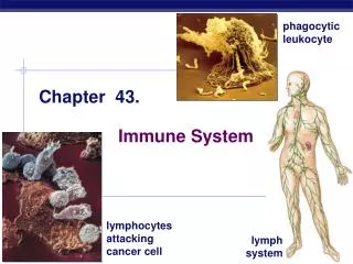
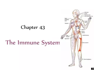
Chapter 43. The Immune System. Overview: Reconnaissance (inspection), Recognition, and Response An animal must defend itself From the many dangerous pathogens it may encounter in the environment or abnormal cells that might develop into cancer

Chapter 43
E N D
Presentation Transcript
Chapter 43 The Immune System
Overview: Reconnaissance (inspection), Recognition, and Response • An animal must defend itself • From the many dangerous pathogens it may encounter in the environment or abnormal cells that might develop into cancer • Two major kinds of defense have evolved that counter these threats • Innate immunity and acquired immunity
3m • Innate immunity • Is present before any exposure to pathogens and is effective from the time of birth • Involves nonspecific responses to pathogens Figure 43.1;a macrophage ingesting a yeast cell.
Innate immune system • nonspecific defense: it does not distinguish between infective agents. • External consisting of epithelial tissue that cover skin and mucus membranes • Internal it get trigered by chemical signals and involves macrophages and other phagocytic ells.
Acquired immunity, also called adaptive immunity • Develops only after exposure to inducing agents such as microbes, toxins, or other foreign substances • Involves a very specific response to pathogens • The recognition involves WBC called lymphocytes that produces the humoral (B cells secreting antibodies) and the cellular (T cells such as cytotoxic cells that directly kill the microbe).
INNATE IMMUNITY Rapid responses to a broad range of microbes ACQUIRED IMMUNITY Slower responses to specific microbes External defenses Internal defenses Skin Phagocytic cells Humoral response (antibodies) Mucous membranes Antimicrobial proteins Secretions Inflammatory response Invading microbes (pathogens) Cell-mediated response (cytotoxic lymphocytes) Natural killer cells Figure 43.2 A summary of innate and acquired immunity
Concept 43.1: Innate immunity provides broad defenses against infection • A pathogen that successfully breaks through an animal’s external defenses • Soon encounters several innate cellular and chemical mechanisms that impede its attack on the body
External Defenses • Intact skin and mucous membranes • Form physical barriers that bar the entry of microorganisms and viruses • Certain cells of the mucous membranes produce mucus • A viscous fluid that traps microbes and other particles such as in lungs.
10m Figure 43.3 • In the trachea, ciliated epithelial cells • Sweep mucus and any entrapped microbes upward, preventing the microbes from entering the lungs
Secretions of the skin and mucous membranes • Provide an environment that is often hostile to microbes • Secretions from the skin • Give the skin a pH between 3 and 5, which is acidic enough to prevent colonization of many microbes • Also include proteins such as lysozyme, an enzyme that digests the cell walls of many bacteria • Stomach acidity; kills many bacteria ingested with food, however, some pathogens survive this acidity such as Hepatitis A virus.
Internal Cellular and Chemical Defenses • Internal cellular defenses • Depend mainly on phagocytosis • Phagocytes, types of white blood cells • Ingest invading microorganisms • Initiate the inflammatory response and produce antimicrobial proteins • None phagocytic cells are called natural killers that play a role in the innate immunity
Pseudopodia surround microbes. 1 Microbes Microbes are engulfed into cell. 2 MACROPHAGE Vacuole containing microbes forms. 3 Lysosome containing enzymes Vacuole Vacuole and lysosome fuse. 4 Toxic compounds and lysosomal enzymes destroy microbes. 5 Microbial debris is released by exocytosis. 6 Phagocytic Cells • Phagocytes attach to their prey via surface receptors • And engulf them, forming a vacuole that fuses with a lysosome Figure 43.4
When engulfing any bacteria or virus, the engulfed mateial fuses with lysozymes which kills by two ways; • Generates toxic forms of oxygen as the super oxide anion and nitric oxide. • Lysosomal enzymes including lysozyme that digests the engulfed material. • However some pathoges has systems to evade these enzymes or they are naturally resistant to such lysosome as the TB pathogen
Four Types of Phagocytic Cells • Neutrophils; comprise 60-70% of total WBCs. • Attracted by chemical signals, they enter infected tissue by amoeboid movement. So they are atracted by chemotaxis • Only live for few days as they destroy themselves when destroying pathogens. • Macrophages; a specific type of phagocyte • Can be found migrating through the body • Can be found in various organs of the lymphatic system and comprise about 5% of WBC.
Macrophages • Long lived phagocytosis • Most wander through interstitial fluid engulfing bacteria, viruses and cell debris by means of extending pseudopodia that can attach to polysaccharides in the bacterial or viral surface. • Some reside permanently in organs and connective tissues. • Eosinophils;have lower phagocytic activity and are crucial against intracellular parasites such as schistosoma mansoni by secreting enzymes damaging the parasite. • Dendritic cells; can ingest microbes but their major role to stimulate development of acquired immunity.
Interstitial fluid bathing the tissues, along with the white blood cells in it, continually enters lymphatic capillaries. 1 Lymphatic capillary Interstitial fluid Fluid inside the lymphatic capillaries, called lymph, flows through lymphatic vessels throughout the body. 2 Adenoid Tonsil Lymphatic vessels return lymph to theblood via two largeducts that drain intoveins near the shoulders. 4 Lymph nodes Blood capillary Tissue cells Spleen Lymphatic vessel Peyer’s patches (small intestine) Within lymph nodes, microbes and foreign particles present in the circulating lymph encounter macro- phages, dendritic cells, and lymphocytes, which carry out various defensive actions. 3 Appendix Lymphatic vessels Masses of lymphocytes and macrophages Lymph node Figure 43.5 The lymphatic system • Plays an active role in defending the body from pathogens
Antimicrobial Proteins • Numerous proteins function in innate defense • By attacking microbes directly or by impeding their reproduction such as lysozymes.
Complement System • About 30 proteins make up the complement system • Which can cause lysis of invading cells and help trigger inflammation. • The presence of invading microbes triggers a cascade of reactions leads to lysis of invaders • In absence of infection these proteins will be inactive • Interferons; produced by viral infected cells • Provide innate defense against viruses and help activate macrophages • There are two types of interferons; Α and β • Some other lymphocytes produce γ interferon that activates macrophages to produce defensins, another antibmicrobial protein that kills wide range of bacteria.
Inflammatory Response • A localized inflammatory response occurs when there is a damage in a tissue due to physical injury or entry of microorganisms. • dilation of small vessels near the injury increases blood flow to the area (causes redness). • The dilated vessels become more permeable allowing fluids and antimicrobial proteins to move into surrounding tissues resulting in localized edema. • Certain chemical signals are important in initiating inflammatory response:
Histamine is released from injured circulating basophils and mast cells in the connective tissue. Histamine increase dialtion thus increase permiability of nearby capillaries. • Prostaglandins also released from white blood cells and damaged cells. These promote blood flow to the injured area and enhances the migration of phagocytic cells from blood into the injury site. • This increased blood flow delivers clotting elements that help block the spread of pathogens and begin the repair process. • Phagocytes are attracted to the damaged tissues by several chemical signals including small proteins called Chemokines.
Neutrophils arrive first, followed by monocytes which develop into phagocytes. • Neutrophils eliminate pathogens and then die. • Macrophages destroy pathogens and clean up the remains of damaged tissues. • In sever infections, e.g. meningitis, • the bone marrow may be stimulated to release more neutrophils by molecules emitted by injured cells and several fold leukocytes will be produced within hours.
A fever may develop in response to toxins released by pathogens, or due to pyrogens released by certain leukocytes. The pyrogens sets the body thermostat at higher temperature. • Moderate fever inhibits the growth of some microorganisms and also facilitate phagocytosis and speed up tissue repair. However, sever fever is dangerous • Certain pathogenes can induce an overwhelming immune response causing what is called septic shock that is characterized by high fever and low blood pressure.
Blood clot Pin Pathogen Macrophage Blood clotting elements Chemical signals Phagocytic cells Phagocytosis Capillary Red blood cell Fluid, antimicrobial proteins, and clotting elements move from the blood to the site. Clotting begins. Chemical signals released by activated macrophages and mast cells at the injury site cause nearby capillaries to widen and become more permeable. Neutrophils and macrophages phagocytose pathogens and cell debris at the site, and the tissue heals. Chemokines released by various kinds of cells attract more phagocytic cells from the blood to the injury site. 1 4 2 3 Major events in the local inflammatory response Figure 43.6
Natural Killer Cells • Natural killer (NK) cells • Patrol the body and attack virus-infected body cells and cancer cells • it is attached to a virus or a cancers cell due to surface receptors on them. • The attachment trigger the release of ceratin chemicals that cause apoptosis in the cells they attack
InvertebrateImmune Mechanisms • Many invertebrates defend themselves from infection • By many of the same mechanisms in the vertebrate innate response • Fruit fly have similar mechanism to vertebrates, they contain hemocytes as equavlent to blood. • Invertbrates lacks lymphocytes, thus they depend heavily on innate immune mechanisms to defend themselves against invaders.
Concept 43.2: In acquired immunity, lymphocytes provide specific defenses against infection • Acquired immunity • Is the body’s second major kind of defense • Involves the activity of lymphocytes
Antigen- binding sites Epitopes (antigenic determinants) Antibody A Antigen Antibody B Antibody C The Antigen Concept • An antigen is any foreign moleculethat is specifically recognized by lymphocytes and elicits a response from them • A lymphocyte actually recognizes and binds to just a small, accessible portion of the antigen called an epitope or an antigenic determinant (4-5 a.a). Figure 43.7
Antigen Recognition by Lymphocytes • The vertebrate body is populated by two main types of lymphocytes • B lymphocytes (B cells) and T lymphocytes (T cells) which circulate through the blood • B and T cells recognize antigens by means of antigen specific receptors in their plasma membranes. • The plasma membranes of both B cells and T cells have about 100,000 antigen receptors that all recognize the same epitope
Antigen- binding site Antigen- binding site Disulfide bridge V V V V Variable regions Light chain C C Constant regions C C Transmembrane region Plasma membrane Heavy chains B cell Cytoplasm of B cell A B cell receptor consists of two identical heavy chains and two identical light chains linked by several disulfide bridges. (a) B Cell Receptors for Antigens • B cell receptors bind to specific, intact antigens • Are often called membrane antibodies or membrane immunoglobulins and are similar to secreted antibodies Figure 43.8a
Antigen- Binding site Variable regions Constant regions Transmembrane region b chain Plasma membrane a chain Disulfide bridge T cell Cytoplasm of T cell (b) • A T cell receptor consists of one • chain and one b chain linked by a disulfide bridge. T Cell Receptors for Antigens and the Role of the MHC • Each T cell receptor consists of two different polypeptide chains V V C C Figure 43.8b
T cells bind to small fragments of antigens • That are bound to normal cell-surface proteins called MHC molecules • MHC molecules • Are encoded by a family of genes called the major histocompatibility complex
Infected cells produce MHC molecules inside the cell and while they are being exported to cell surface, they bind a fragment of protein from antigens and will present it on the surface. This process is called antigen presentation • A nearby T cell • Can then detect the antigen fragment displayed on the cell’s surface and will elicit the production of a clone of T- cells that are specific for this particular peptide.
Depending on their source • Peptide antigens are handled by different classes of MHC molecules
Class I MHC molecules • Found on almost all nucleated cells of the body • Display peptide antigens to cytotoxic T cells • Infected and cancerous cells display such peptide antigens.
Infected cell 1 Antigen fragment 1 A fragment of foreign protein (antigen) inside the cell associates with an MHC molecule and is transported to the cell surface. Class I MHC molecule 2 T cell receptor 2 The combination of MHC molecule and antigen is recognized by a T cell, alerting it to the infection. (a) Cytotoxic T cell Class I MHC molecules 1 Figure 43.9a
Class II MHC molecules • Located mainly on dendritic cells, macrophages, and B cells (Antigen presenting cells, APC). • Bind peptides derived from foreign materials that have been internalized by phagocytosis. • Display antigens to helper T cells • Humans have large number of different MHC alleles in human population. • Any two people (except identical twins) are very unlikely to have same set of MHC molecules. • MHC provides fingerprint of each individual
1 Microbe Antigen- presenting cell 1 A fragment of foreign protein (antigen) inside the cell associates with an MHC molecule and is transported to the cell surface. Antigen fragment 2 2 Class II MHC molecule T cell receptor The combination of MHC molecule and antigen is recognized by a T cell, alerting it to the infection. Helper T cell (b) Class II MHC molecules Figure 43.9b
Lymphocyte Development • Lymphocytes • Arise from pluripotent stem cells in the bone marrow • Newly formed lymphocytes are all alike • But they later develop into B cells or T cells, depending on where they continue their maturation. • There are three events in the life of a lymphocyte • First 2 events are;Lymphocyte maturation of B or T • Third event is the lymphocyte encounter with an antigen that leads to its activation, proliferation and differentiation, a process called clonal selection.
Bone marrow Lymphoid stem cell Thymus T cell B cell Blood, lymph, and lymphoid tissues (lymph nodes, spleen, and others) Figure 43.10 Development and Differentiation of lymphocyte Both circulate in
Generation of Lymphocyte Diversity by Gene Rearrangement • B and T lymphocytes recognize specific epitopes on antigens via their antigen receptors. • A single B or T cell has about 100,000 or these receptors. • The variable region at the tip of each antigen receptor chain (antigen binding site) forms this diversity. • The sequence of a.a. in these regions varies from cell to cell, thus it is estimated that each individual has as many as 1 million different B cells and 10 million T-cells each respond to different antigen. • Early in development, random, permanent gene rearrangement Forms functional genes encoding the different B or T cell antigen receptor chains.
V4–V39 3 2 4 1 DNA of undifferentiated B cell V40 J1 J2 J3 J4 J5 V2 V3 Intron V1 C Deletion of DNA between a V segmentand J segment and joining of the segments DNA of differentiated B cell V3 V1 V2 J5 Intron C Transcription of resulting permanently rearranged,functional gene V3 J5 C pre-mRNA Intron RNA processing (removal of intron; addition of capand poly (A) tail) V3 J5 Cap C Poly (A) mRNA Translation V C Light-chain polypeptide B cell receptor Variable region Constant region B cell Figure 43.11 Immunoglobulin gene rearrangement
Summary of the immunoglobulin gene rearrangement • Immunglobulin light chain gene contains a series of 40 variable gene segments separated by long stretch of DNA from the 5 Joining (J) gene segments. • Down stream of the J segments there is an intron and an exon (C) that codes for the constant region. • Early in development, a set of enzymes called recombinase links one V gene to a J segment eliminating the long stretch of DNA and forming one exon • Recombinase acts randomly thus it could join any of the V gene segments to the J segments i.e 200 combinations with only one combination/cell. • Once this combination takes place, the gene can be transcribed mRNA processed and translated to a light chain with both variable and constant regions. • The light chain produced will randomly combine with a heavy chain that was made in the same way.
Testing and Removal of Self-Reactive Lymphocytes • As B and T cells are maturing in the bone and thymus • Their antigen receptors are tested for possible self-reactivity • Lymphocytes bearing receptors for antigens already present in the body • Are destroyed by apoptosis or rendered nonfunctional • The immune system exhibits the critical feature of self-tolerance.
Clonal Selection of Lymphocytes • Binding of antigen to a mature lymphocyte induces the lymphocyte’s proliferation and differentiation, a process called clonal selection. • Clonal selection generates; • a clone of short-lived activated effector cells • a clone of long-lived memory cells
Antigen molecules bind to the antigen receptors of only one of the three B cells shown. Antigen molecules B cells that differ in antigen specificity Antigen receptor The selected B cell proliferates, forming a clone of identical cells bearing receptors for the selecting antigen. Some proliferating cells develop into short-lived plasma cells that secrete antibodies specific for the antigen. Some proliferating cells develop into long-lived memory cells that can respond rapidly upon subsequent exposure to the same antigen. Antibody molecules Clone of memory cells Clone of plasma cells Figure 43.12 Clonal selection of B cells
The nature and function of the effector cell depends on whether the lymphocyte selected is a helper T cell, a cytotoxic T cell or a B cell. • The other clone consist of memory cells that live long and bear receptors for specific for same inducing antigen. • So the clonal selection can be defined as this; each antigen by binding to specific receptor, selectively activates a tiny fraction of cells from the body’s diverse pool of lymphocytes that is dedicated to destroy the antigen.
Primary Immune Response • Is the proliferation of lymphocytes to form effecter cells specific to an antigen during the body’s first exposure to the antigen. • There is 10-17 days lag period between initial exposure and maximum production of effecter cells. Mainly IgM • The lympnocytes selected by the antigen are differentiated into B and T cells during the lag period. • Activated B cells give rise to effecter cells called plasma cells which secrete antibodies (humoral response).
1 Secondary response to anti- gen A produces antibodies to A; primary response to anti- gen B produces antibodies to B 4 Day 1: First exposure to antigen A 2 Primary response to antigen A produces anti- bodies to A 3 Day 28: Second exposure to antigen A; first exposure to antigen B 104 103 Antibody concentration (arbitrary units) 102 Antibodies to A Antibodies to B 101 100 35 28 21 42 49 56 0 14 7 Figure 43.13 Time (days) Secondary immune response • Memory cells facilitate a faster, more efficient response that takes 2-7 days. IgG mainly
Secondary response leads to the production of very high amount of antibodies that have more affinity than the antibodies produced in the primary response. • The immunes system capacity to generate secondary immune response is called Immmunological memory
Concept 43.3: Humoral and cell-mediated immunity defend against different types of threats • Acquired immunity includes two branches • The humoral immune response involves the activation and clonal selection of B cells, resulting in the production of secreted antibodies • The cell-mediated immune response involves the activation and clonal selection of cytotoxic T cells