Download
1 / 38
390 likes | 746 Vues
Chapter 11 How Cells Reproduce. 11.1 Henrietta ’ s Immortal Cells. Henrietta Lacks died of cancer in 1951, at age 31, but her cells ( HeLa cells) are still growing in laboratories

E N D
11.1 Henrietta’s Immortal Cells • Henrietta Lacks died of cancer in 1951, at age 31, but her cells (HeLa cells) are still growing in laboratories • HeLa cells are widely used to investigate cancer, viral growth, protein synthesis, the effects of radiation, and other processes important in medicine and research • Understanding why cancer cells are immortal – and why we are not – begins with understanding the structures and mechanisms that cells use to divide
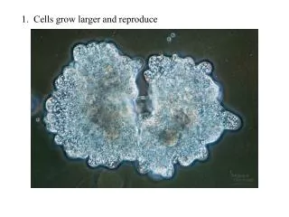
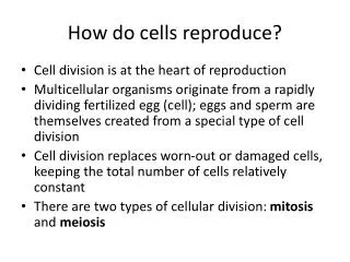
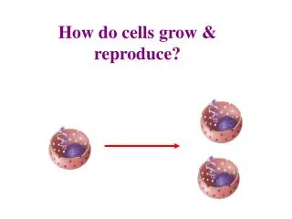
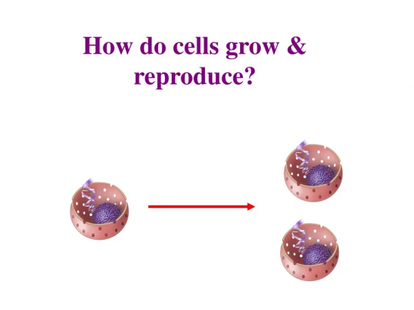
11.2 Multiplication by Division • Division of a eukaryotic cell typically occurs in two steps: nuclear division followed by cytoplasmic division • The sequence of stages through which a cell passes during its lifetime is called the cell cycle
Interphase • Interphase consists of three stages, during which a cell increases in size, doubles the number of cytoplasmic components, and replicates its DNA • G1: Interval of cell growth and activity • S: Interval of DNA replication (synthesis) • G2: Interval when the cell prepares for division
Interphase and the Life of a Cell • Most cell activities take place during G1 • Control mechanisms work at certain points in the cell cycle; some can keep cells in G1 • Loss of control may cause cell death or cancer
Mitosis and Asexual Reproduction • Mitosis is a nuclear division mechanism that maintains the chromosome number • In multicelled species, mitosis and cytoplasmic division increase cell number during development , and replace damaged or dead cells later in life • In asexual reproduction, a single individual can reproduce by mitosis and cytoplasmic division
Homologous Chromosomes • Homologous chromosomesare pairs of chromosomes having the same length, shape, and genes • Typically, one member of a homologous pair is inherited from the mother, the other from the father • Human cells have 46 chromosomes (23 pairs) • Except for a pairing of sex chromosomes (XY) in males, the chromosomes of each pair are homologous
Chromosomes During Division • A cell can’t function without a full complement of DNA – each cell needs to have one copy of each chromosome • The cell replicates its DNA in preparation for mitosis • Four stages of mitosis (prophase, metaphase, anaphase, and telophase) parcel sister chromatids into separate nuclei • Cytoplasmic division results in two diploid cells, each with the same number and kind of chromosomes as the parent
A An unduplicated pair of chromosomes in a cell in G1. B By G2, each chromosome has been duplicated. C Mitosis and cyto-plasmic division package one copy of each chromosome into each of two new cells. Stepped Art Figure 11-4 p179
Controls Over the Cell Cycle • Most differentiated human cells are in G1 – some types never progress past that stage • When a cell divides is determined by gene expression controls; some cause the cell cycle to advance, others stop the cycle from proceeding • These built-in checkpoints allow problems to be corrected before the cycle proceeds
interphase prophase G2 metaphase S anaphase mitosis telophase G1 Figure 11-2 p177
Take-Home Message: What is cell division and why does it occur? • The sequence of stages through which a cell passes during its lifetime (interphase, mitosis, and cytoplasmic division) is called the cell cycle • A eukaryotic cell reproduces by division: nucleus first, then cytoplasm; each descendant cell receives a set of chromosomes and some cytoplasm • Mitosis is the basis of body size increases and tissue repair in multicelled eukaryotes; it is also the basis of asexual reproduction in single-celled and some multicelled eukaryotes • Gene expression controls advance, delay, or block the cell cycle in response
Preparing for Mitosis • During interphase, a cell’s chromosomes are loosened to allow transcription and DNA replication • In preparation for nuclear division, chromosomes condense into their most compact “X” forms • Tight condensation keeps the chromosomes from getting tangled and breaking during nuclear division
Interphase in Plant and Animal Cells Onion root cell Whitefish embryo cell
Take-Home Message: What is the sequence of events in mitosis? • Each chromosome in a cell’s nucleus was duplicated before mitosis begins, so each consists of two DNA molecules • Mitosis proceeds in four stages: prophase, metaphase, anaphase, and telophase • During these four stages, sister chromatids of the duplicated chromosomes are separated and packaged into two new nuclei; each new nucleus contains the same number and types of chromosomes as the parent cell
11.4 Cytokinesis: Division of Cytoplasm • In most kinds of eukaryotes, the cell cytoplasm divides between late anaphase and the end of telophase, but the mechanism of division differs between plants and animals • Cytokinesis • The process of cytoplasmic division
Cytokinesis in Animal and Plant Cells • Animal cells • A cleavage furrow partitions the cytoplasm • A band of actin filaments rings the cell midsection, contracts, and pinches the cytoplasm in two • Plant cells • A cell plateforms midway between the spindle poles; it partitions the cytoplasm when it reaches and connects to the parent cell wall
Cytoplasmic Division in Animal Cells After mitosis is completed, the spindle begins to disassemble. 1 At the midpoint of the former spindle, a ring of actin and myosin filaments attached to the plasma membrane contracts. 2 This contractile ring pulls the cell surface inward as it shrinks. 3 The ring contracts until it pinches the cell in two. 4
Cytoplasmic Division in Plant Cells The future plane of division was established before mitosis began. Vesicles cluster here when mitosis ends. 5 As the vesicles fuse with each other, they form a cell plate along the plane of division. 6 The cell plate expands outward along the plane of division. When it reaches the plasma membrane, it attaches to the membrane and partitions the cytoplasm. 7 The cell plate matures as two new cell walls. These walls join with the parent cell wall, so each descendant cell becomes enclosed by its own cell wall. 8
Take-Home Message:How do cells divide? • After mitosis, the cytoplasm of the parent cell typically is partitioned into two descendant cells, each with its own nucleus • In animal cells, a contractile ring pinches the cytoplasm in two • In plant cells, a cell plate that forms midway between the spindle poles partitions the cytoplasm when it reaches and connects to the parent cell wall
11.5 Marking Time With Telomeres • Telomeres protect eukaryotic chromosomes from losing genetic information at their ends • Shortening telomeres are associated with aging
Telomeres • The ends of eukaryotic DNA strands consist of noncoding sequences called telomeres • Vertebrate telomeres have a short DNA sequence, 5′-TTAGGG-3′, repeated thousands of times • Telomeres are important because a polymerase can’t copy the last hundred or so bases of the 3′ end of a chromosome • When telomeres get too short, checkpoint gene products stop the cell cycle, and the cell dies
Stem Cells and Telomerase • In an adult, only stem cells retain the ability to divide indefinitely • Stem cells are immortal because they retain the ability to make telomerase, a molecule that reverses telomere shortening that normally occurs after DNA replication • Research suggests that the built-in limit on cell divisions may be part of the mechanism that sets an organism’s life span
Take-Home Message:What is the function of telomeres? • Telomeres at the ends of chromosomes provide a buffer against loss of genetic information • Telomeres shorten with every cell division in normal body cells; when they get too short, the cell stops dividing and dies
Checkpoint Failure and Tumors • When enough checkpoint mechanisms fail, a cell loses control over its cell cycle forms a neoplasm – a group of cells that lost control over how they grow and divide • A neoplasm that forms a lump (abnormal mass) in the body is called a tumor
Oncogenes and Proto-Oncogenes • An oncogene is any gene that helps transform a normal cell into a tumor cell • Genes encoding proteins that promote mitosis are called proto-oncogenes– mutations can turn them into oncogenes • Growth factors are molecules that stimulate a cell to divide and differentiate • A gene that encodes the epidermal growth factor (EGF) receptor is an example of a proto-oncogene
Tumor Suppressors • Checkpoint gene products that inhibit mitosis are called tumor suppressors because tumors form when they are missing • The products of the BRCA1 and BRCA2 genes are examples of tumor suppressors – they regulate the expression of DNA repair enzymes
Three Characteristics of Cancer Cells • Cancer cells grow and divide abnormally; capillary blood supply to the cells may increase abnormally • Cytoplasm and plasma membrane are altered; malignant cells typically have an abnormal chromosome number, and metabolism may shift toward fermentation • Cancer cells have altered recognition proteins and weakened adhesion – malignant cells break loose and invade other parts of the body (metastasis)
Cells of a malignant neoplasm can break away from their home tissue. Benign neoplasms grow slowly and stay in their home tissue. 2 1 3 The malignant cells become attached to the wall of a blood vessel or lymph vessel. They release digestive enzymes that create an opening in the wall, then enter the vessel. The cells creep or tumble along inside blood vessels, then leave the bloodstream the same way they got in. They often start growing in other tissues, a process called metastasis. 4 Figure 11-11 p185
Reducing the Risk of Cancer • Each year, cancer causes 15 to 20 percent of all human deaths in developed countries • Life style choices such as not smoking and avoiding exposure of unprotected skin to sunlight can reduce the risk of acquiring mutations that lead to cancer • Some neoplasms can be detected with periodic screening procedures such as Pap tests or dermatology exams
Skin Cancers A Basal cell carcinoma is the most common type of skin cancer. This slow- growing, raised lump may be uncolored, reddish- brown, or black. B The second most common form of skin cancer is a squamous cell carcinoma. This pink growth, firm to the touch, grows under the surface of skin.
Take-Home Message:What is cancer? • Cancer is a disease that occurs when the abnormally dividing cells of a neoplasm physically and metabolically disrupt body tissues • A malignant neoplasm results from mutations in multiple checkpoint genes • Although some mutations are inherited, life style choices and early intervention can reduce one’s risk of cancer