Download
1 / 38
380 likes | 665 Vues
Venous Thromboembolic Disease: Genetics and Treatment. John P. Sheehan, M.D. Section of Hematology University of Wisconsin Madison, WI. Venous Thromboembolic Disease (VTE): Scope of the Problem. 1) 250,000-300,000 cases annually in U.S. 2) Overall annual incidence ~1/1000

E N D
Venous Thromboembolic Disease:Genetics and Treatment John P. Sheehan, M.D. Section of Hematology University of Wisconsin Madison, WI
Venous Thromboembolic Disease (VTE):Scope of the Problem 1) 250,000-300,000 cases annually in U.S. 2) Overall annual incidence ~1/1000 3) Prevalence rises with age: from 1/100,000 to 1/100 in the elderly 4) Complications include: a) 1-2% incidence of fatal PE b) 20-30% post-thrombotic syndrome c) 10-35% recurrence rates after therapy
Natural History of Acute DVT(Pradoni, Ann Intern Med, 1996) 1) 355 consecutive pts with first DVT (pop. 350,000). 2) Therapy: heparin (10 days) + warfarin (3 months). 3) Recurrent VTE- 78 pts, 24.6% at 5 yr a) 44.9% ipsilateral, 35.9% contralateral DVT b) 19.2% PE, 11.5% fatal (7/9 dx @ autopsy) c) Under diagnosis of PE likely in this cohort. 4) Post-thrombotic syndrome- 28 % total, 9.8% “severe”.
Risk Factors for Recurrent VTE(Pradoni, Ann Intern Med, 1996)
Benefit of Heparin Therapy A) Brandjes et al., NEJM, 1992 1) Heparin + acenocoumarol vs. acenocoumarol (OAC only) in patients with first DVT w/o PE. 2) Terminated early due to excess events (symptomatic extension/recurrent VTE) in OAC only group 3) 4/60 (6.7%) combined therapy vs. 12/60 (20%) OAC only 4) Asymptomatic extension/PE in first week: 8.2% vs. 39.4% B) Hull et al., NEJM, 1990 5 days equivalent to 10 days of heparin therapy (3 months)
Benefit of Oral Anticoagulant Therapy 1) Warfarin vs. low dose heparin, 4 vs. 25% recurrence rate in first 3 months (Hull, NEJM, 1979) 2) DURAC I Trial- 902 pts, 1st DVT (Schulman, NEJM, 1995) a) 2 yr recurrence: 9.5% (6 months) vs. 18.1%(6 wks) b) Exclusions-cancer, PS/PC/AT, > 1 previous VTE 3) DURAC II Trial- 227 pts, 2nd DVT (Schulman, NEJM, 1997) a) 4 yr recurrence: 20.7% (6 months) vs. 2.6%(indefinite) b) Major hemorrhage 2.7 vs. 8.6%worse c) Exclusions: cancer, PS/PC/AT
Heparin Therapy: Risks 1) Risk at therapeutic doses a) major bleeding 1-3% b) fatal bleeding 0-0.8% 2) Clinical risk factors- a) recent surgery/trauma b) co-morbid conditions c) age > 70, ASA/NSAID use 3) Bleeding risk similar for UH and LMWH
Warfarin Therapy: Risks 1) Risk at therapeutic doses a) major bleeding: 3-4%/yr b) fatal bleeding: 0-0.8%/yr c) total bleeding: 8-9%/yr d) ICH: 0.1-0.3%/yr, age > 60: 0.3-1.0%/yr 2) Clinical risk factors- a) INR (intensity of anticoagulation) b) age >65 c) co-morbid conditions, hx of GI/GU bleeding, CVA d) first month of therapy
VTE: Clinical Questions 1) When does the benefit of antithrombotic therapy outweigh the risk of bleeding complications? 2) Can we adequately predict the risk of VTE to determine who will benefit from prolonged therapy?
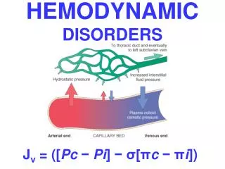
Pathogenesis of VTE A) Multicausal disease 1) Acquired factors- “clinical milieu” 2) Genetic risk factors- multigenic etiology B) Virchow’s triad- 1) venous stasis 2) endothelial/vessel injury 3) intrinsic hypercoagulability
Acquired Risk Factors for VTE 1) surgery/general anesthesia/trauma 2) malignancy (and chemotherapy), MPD 3) immobilization/lower extremity cast/obesity 4) pregnancy (esp. post-partum) 5) estrogens (OC and HRT) 6) CHF/venous insufficiency/obstruction 7) lupus anticoagulant/antiphospholipid Ab 8) heparin-induced thrombocytopenia
Treatment of “Idiopathic” VTE(Kearon, NEJM, 1999) 1) Exclusions: a) fx/cast/immobilized x 3 d/general anesthesia (3 months) b) cancer (5 yrs) c) known AT/PC/PS deficiency (not tested) d) contraindication to OAC, antiplatelet drugs, pregnant 2) Randomized at 3 months to placebo or continued OAC. 3) Terminated early due to excess recurrent VTE in placebo arm (27.4 vs. 1.3% recurrences/pt-yr). 4) Bleeding increased in OAC arm (total 1.4 vs. 11.5 %).
Idiopathic VTE: Duration of Therapy (Agnelli et al, NEJM, 2001) 1 ) 267 consecutive patients w/ 1st episode idiopathic DVT 2 ) Excluded cancer, known thrombophilia, immobilization, trauma, surgery, childbirth, pregnancy, OCP use, other indication for OAC, or life expectancy less than 2 years. 3) Randomized after 3 months warfarin (INR 2-3) to additional 9 months or no further therapy. 4) Intent-to-treat outcomes after 2 yr. followup: a) VTE recurrence: 15.7% (9 mo.) vs. 15.8% (all idiopathic) b) Major Bleeding: 4/134 vs. 2/133
Multigenic Etiology of VTE 1) Gene defect ≠ Disease Ex: PC deficiency is a risk factor, not a disease 2) The most common gene defects are only present in a minority of VTE cases. Ex: FV Leiden present in 20-40% of VTE cases 3) Multiple defects interact, often synergistically. Ex: FV Leiden and PC deficiency 4) Ascertainment bias profoundly affects relative risk estimates for specific gene defects. Ex: general population vs. thrombophilic family
Recurrent VTE Risk- Effect of Factor V Leiden (FVL) and PT 20210A 1) Majority of studies do not demonstrate increased risk for patients with isolated FVL or PT 20210A mutations. 2) Why do single gene defects not consistently predict increased recurrence risk? a) selection- patients with acquired risk factors (ie. cancer) excluded to variable extent, enriching for idiopathic cases (with multiple gene defects-known and unknown). b) failure to account for ethnicity in the study population. 3) In contrast, combined FVL/PT20210A mutations consistently predict increased risk of recurrent VTE.
Gene-Environment Interactions in VTE(Martinelli et al, Arteriosclero Thromb Vasc Biol, 1999)
Genetic Risk Assessment for VTE 1) How many genes are involved? 2) What is the contribution of known vs. unknown genes? 3) Are specific gene-gene interactions synergistic, additive, or have overlapping functions with regard to VTE risk? 4) Difficulties: a) incomplete database b) population differences (ethnicity)
Clinical Assessment for VTE 1) History of previous thrombotic events? (Documentation?) 2) Family history of thrombosis? 3) Age at first event? 4) Unusual location or frequency of thrombosis? Ex: mesenteric venous or cerebral sinus thrombosis 5) Risk Factor Assessment a) transient/reversible clinical factors b) ongoing risk factors i) clinical factors ii) genetic defects
Impact on Treatment Decisions A) Problem: How should we treat idiopathic VTE? 1) Risk assessment expressed in terms of relative risk. 2) Cost of therapy is known in terms of absolute risk. 3) Rationale treatment decisions require comparison of absolute risks over time (include mortality data). B) Strategies for conflict resolution 1) Risk stratification- only expose patients at greatest risk to chronic anticoagulation. 2) Improve risk/benefit ratio of antithrombotic therapy.
Optimizing Warfarin (OAC) Management 1) INR to correct for thromboplastin sensitivity 2) Anticoagulation clinics 3) Point-of-care testing 4) Patient self-management
Alternative Therapy for VTE • 1) Low molecular weight heparin • 2) Heparin pentasaccharide • 3) Oral heparin- SNAC complex • 4) Oral thrombin inhibitor- Ximegalatran • 5) Experimental targets- • a) TF-FVIIa- nematode anticoagulant protein • b) FIXa-FVIIIa- “low affinity” heparanoids • c) exosite-directed inhibitors
TF VIIa IXa VIIIa Xa Va Thrombin XI IX Injury XIa VIII Initiation X V PT Xa Propagation Fibrin Fibrinogen
Heparin: Mechanisms of Action A) Antithrombin dependent 1) Inhibition of factor IXa, Xa, and thrombin 2) Inhibition of factor VIIa-tissue factor B) Antithrombin independent 1) Inhibition of factor VIII activation by thrombin 2) Inhibition of the intrinsic tenase complex 3) Anti-platelet/anti-adhesion properties
Low Molecular Weight Heparin 1) Equivalent clinical efficacy to UH for DVT/PE. 2) Risks- HIT lower, bleeding similar to UH. Need to recognize potential for spinal/epidural hematoma. 3) Important features- a) retains heterogeneity of UH ( chain length). b) anti-factor Xa/anti-IIa ratio (w/o CaCl2 ) c) does not reliably prolong the APTT 4) Major Advantage: improved pharmacokinetics (predominantly renal elimination).
Fondaparinux (Org 31540/SR90107A) 1) Synthetic pentasaccharide-binds ATIII with high affinity. 2) T1/2 ~ 15-20 hours, ~100% bioavailable SQ, renal clearance. Minimal to no prolongation of APTT. 3) Bleeding risk increased w/ renal insufficiency, body weight < 50 kg, or age > 70 y/o. No antidote. 4) Risks: spinal/epidural hematoma likely similar to LMWH, HIT risk decreased? 5) Approval for DVT prophylaxis in knee/hip arthroplasty.
Low Affinity Heparin (LAH) 1) Depleted of high affinity pentasaccharide. 2) Retains significant antithrombotic efficacy in rabbit venous thrombosis model, minimal effect on bleeding. 3) Inhibits thrombin generation in plasma. 4) Inhibits intrinsic tenase (fIXa-fVIIIa) activity at therapeutic heparin concentrations.
Unfractionated Heparin (UH) Inhibits the Intrinsic Tenase Complex
Heparin Oligosaccharide Allosteric Effect FIXa FX Protease FVIIIa
Conclusions-I 1) Evaluation of clinical risk factors is critical to assessing the likelihood of VTE recurrrence. 2) Idiopathic VTE has a strong genetic component and higher recurrence rate, representing multiple gene defects. 3) Current assessment of genetic component limited by incomplete data base. 4) Improved VTE management will require: a) comprehensive, rapid genetic screens b ) therapies with improved safety for chronic usage.
Conclusions-II 5) Improved genetic risk assessment: a) use of multiple SNP patterns in candidate genes b) identification of novel genes in defined populations or phenotypic screens in vertebrate animal models. c) proteomic analysis of plasma and/or platelet factors. 6) Improved drug therapy: a) alternative enzyme complexes (FIXa-FVIIIa or TF- FVIIa) and molecular (exosite-directed) targets. b) increased safety for broader patient population. c) will reduce importance of genetic risk assessment.