Download
1 / 26
260 likes | 283 Vues
Join the Retina Imaging Conference with Dr. Brett Mueller at the University of Louisville. Explore a patient case of Central Retinal Vein Occlusion, its risk factors, evaluation, management, and the latest treatment strategies. Discover the CRUISE study outcomes and insights on CRVO in young patients.

E N D
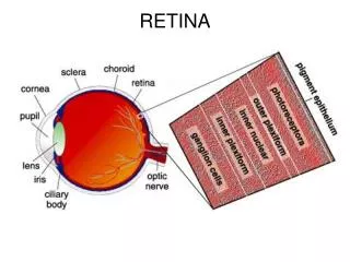
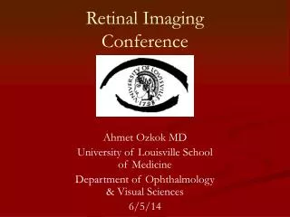
Retina Imaging Conference Brett Mueller, D.O., Ph.D. 9/10/2015 University of Louisville Department of Ophthalmology and Visual Sciences
Patient Presentation • CC: Decreased Vision Right Eye • HPI: 38 yo WM presents w/ blurry vision OD for one week. Pt states it happened suddenly w/out pain. Pt denies any history of trauma or straining. Of note, the patient is an avid weightlifter that uses supplements including ephedera (dietary supplement containing ephedrine and pseudoephedrine).
History POHx: none PMHx: Hypertension w/ most recent PCP visit being 178/81. He stated he is not taking any blood pressure medications at this time. FAMHx: none ROS: none MEDS: none ALLERGIES: none
18 3→2 20/80+1 (+1.25 sph) 21 3→2 20/20 Exam VA TP P no RAPD EOM: full OU CVF: full OU
Exam OD OS LIDS/LASHES WNL WNL CONJ WNL WNL CORNEA WNL WNL IRIS WNL WNL LENS WNLWNL
Fundus Photos OD OS OD photo demonstrates pre-retinal and intraretinal hemorrhages extending along the arcades and in the macula
FAF Photos OD OS OD photo demonstrates pre-retinal and intraretinal hemorrhages extending along the arcades and in the macula
1.OD 2.OD Macula is flat with no evidence of CME. Pre-retinal, sub-ILM blood demonstrated through the retinal hemorrhages.
FA Early AV Phase Arterial Phase 00:20:00 00:13:00 Early arterial phase shows good arterial filling w/ no ischemic retinal areas. Early AV phase demonstrates venous laminar filling.
FA AV Phase AV Phase 00:40:00 00:30:00 Both AV phase photos at 30 and 40 seconds demonstrate delayed filling of the venous circulation w/ prolonged laminar flow.
Labs • WBC: 5.2 (4.5-10) • Hemoglobin: 14.6 (13.5-17.5) • Hematocrit: 44.2 (38.8-50) • Platelets: 250 (150-450) • Fibrinogen: 236 (212-476) • PT: 13.6 (11.4-15.1) • PTT: 27.2 (21.1-38.4)
Labs • Protein C: 86 (70-180) • Protein S: 123 (70-150) • Antiphospholipid antibody: Anticardiolipin, Beta-2 glycoprotein, antiphospholipid antibody and lupus anticoagulation: negative • Factor V Leiden: negative
Summary 38 y/o WM with decreased vision OD. Examination and FA reveal multiple pre-retinal and intraretinal hemorrhages with delayed venous filling. Patient has a history of weight lifting and supplement consumption that contain ephedrine and pseudophedrine. DDx: • Non-ischemic Central Retinal Vein Occlusion • Valsalva Retinopathy PLAN: OBSERVATION with 1 month f/u
11 4→2 20/CF 2ft (+1.25 sph) 12 4→2 20/20 Exam 1 month later VA TP P SC no RAPD EOM: full OU
1 month later OD OS OCT: OD demonstrates serous neurosensory retinal detachment
Central Retinal Vein Occlusion • Disease of the arterial circulation • Divided into non-ischemic and ischemic disease • Mechanism of action: Thrombosis of the central retinal vein at or posterior to the level of the lamina cribrosa.
Risk Factors for Central Retina Vein Occlusion • Age (90% greater than 50), systemic arterial hypertension, open-angle glaucoma, diabetes mellitus and hyperlipidemia. • Oral contraceptives and diuretics have also been associated as risk factors for the development of CRVO
Risk Factors for Central Retina Vein Occlusion • Rare predisposing hypercoagulable conditions associated with this disease include: • Hyperhomocysteinemia • Protein S deficiency • Protein C deficiency • Disorders associated with vasculitis like sarcoidosis and lupus.
Central Retinal Vein Occlusion Evaluation and Management • Determination should be made if the patient has non-ischemic or ischemic CRVO. • Absence of treatment, patients with CRVO should be monitored monthly for the first 6 month for the development of anterior segment neovascularization. • Most common complications include vitreous hemorrhage, anterior segment neovascularization, and neovascular glaucoma.
Central Retinal Vein Occlusion • Treatment • CRUISE (Study of the Efficacy and Safety of Ranibizumab Injection in Patients With Macular Edema Secondary to CRVO) • Phase III Multicenter, randomized, controlled clinical trial 392 eyes with CRVO and secondary macular edema (>250μm), BCVA of 20/40 to 20/320 • 3 groups: Sham injection • Ranibizumab (Lucentis) 0.3mg • Ranibizumab 0.5mg
Central Retinal Vein Occlusion Treatment • CRUISE (Study of the Efficacy and Safety of Ranibizumab Injection in Patients With Macular Edema Secondary to CRVO) • Results: At month 6, BCVA gains of >15 ETDRS letters in 46.2% of patients receiving 0.3 mg, 47.7% of those receiving 0.5 mg, and 16.9% of those receiving sham injections. • GALILEO and COPERNICUS Demonstrated similar effects but with aflibercept.
Central Retinal Vein Occlusion in the Young • Anomalous retinal anatomy at the level of the optic nerve • Hyperviscosity syndromes caused by autoimmune diseases or cancers (usually bilateral) • Antiphospholipid syndrome, increased homocysteine, Factor C/S def. • Malignant hypertension w/ CRF
Case report identifying 2 healthy, athletic young patients (< 40 yo) that developed a CRVO and had a negative hypercoagulation workup. • Both patients had complete resolution of their ME and returned to 20/20 vision after getting 3 monthly intravitreal injections of Avastin. Moisseiev E, Sagiv O, Lazar M. Intense exercise causing central retinal vein occlusion in a young patient: case report and review of the literature. Case Rep Ophthalmol. 2014 Apr 5;5(1):116-20. doi: 10.1159/000360904. eCollection 2014 Jan. PubMed PMID: 24847256; PubMed Central PMCID: PMC4025055.
Studied 55 patients younger than 56 years of age (mean age 44) to investigate whether hypercoagulability plays a role in the thrombus formation in patients with a CRVO. • Results • 27% of patients had one positive test result suggesting hypercoagulability. • 4 patients had elevated homocysteine levels • 2 patients had a Factor V Leiden mutation • 3 patients had protein S deficiency. • 3 patients had + lupus anticoagulant • 3 patients had + anticardiolipin antibodies Lahey JM, Tunç M, Kearney J, Modlinski B, Koo H, Johnson RN, Tanaka S. Laboratory evaluation of hypercoagulable states in patients with central retinal vein occlusion who are less than 56 years of age. Ophthalmology. 2002 Jan;109(1):126-31. PubMed PMID: 1177259
References 1. Retina and Vitreous, BSCS 2. Moisseiev E, Sagiv O, Lazar M. Intense exercise causing central retinal vein occlusion in a young patient: case report and review of the literature. Case Rep Ophthalmol. 2014 Apr 5;5(1):116-20. doi: 10.1159/000360904. eCollection 2014 Jan. PubMed PMID: 24847256; PubMed Central PMCID: PMC4025055. 3. Lahey JM, Tunç M, Kearney J, Modlinski B, Koo H, Johnson RN, Tanaka S. Laboratory evaluation of hypercoagulable states in patients with central retinal vein occlusion who are less than 56 years of age. Ophthalmology. 2002 Jan;109(1):126-31. PubMed PMID: 1177259 4. Kanski’s Clinical Ophthalmology A systemic Approach, Eighth Edition. Brad Bowling 5. Gass JDM. Stereoscopic Atlas of Macular Diseases: Diagnosis and Treatment, 4th ed. St. Louis, Mosby, 1997. 6. Barry C, Singh J, Constable IJ. Are optic disc drusen exhibiting Autofluorescence, pseudofluorescence or reflectance? Journal of Ophthalmic Photography 22:32-35, 2000. 7. Thach AB, Yau L, Hoang C, Tuomi L. Time to clinically significant visual acuity gains after ranibizumab treatment for retinal vein occlusion: BRAVO and CRUISE trials. Ophthalmology. 2014 May;121(5):1059-66. doi: 10.1016/j.ophtha.2013.11.022. Epub 2014 Jan 11 8. Kavoussi SC, Kempton JE, Huang JJ. Central retinal vein occlusion resulting from anomalous retinal vascular anatomy in a 24-year-old man. Clin Ophthalmol. 2015 May 20;9:885-7. doi: 10.2147/OPTH.S84214. eCollection 2015. PubMed PMID: 26056427; PubMed Central PMCID: PMC4445948.