Deformities
Deformities. By Prof.Dr./ Lotfy Yones. Professor of Orthopedics Tanta University. Definition. It is a visible abnormality in shape of any part of the body. Causes. A- Congenital : it is seen at birth .may be due to : 1- Chromosomal . 2- Some drugs

Deformities
E N D
Presentation Transcript
Deformities By Prof.Dr./ Lotfy Yones Professor of Orthopedics Tanta University
Definition • It is a visible abnormality in shape of any part of the body .
Causes A- Congenital : it is seen at birth .may be due to : 1- Chromosomal . 2- Some drugs 3- Abnormal intrauterine fetal posture . B- Acquired : 1-Developmental .{genetic , metabolic or hormonal } 2- Traumatic { skin , muscles , joints or bone } 3- Nerve injuries . 4- Inflammation { arthritis , myositis } 5- Bone softening diseases {rickets, osteomalacia etc } 6- Infection .
Diagnosis 1-History taking 2-Laboratory investigations 3-Imaging : a-X-ray b- CT C-MRI
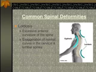
It is a backward angulations above 40 degrees . • Types : I-Mobile • Compensatory { exaggerated lordosis} • Postural {bad habit of sitting in adolescence } • Muscle weakness II-Fixed • Angular {fracture , T.B., Calve disease} • Rounded (regular) {Scheuermann disease , senile kyphosis ,ankylosing spondylitis } • Treatment 1- of the cause 2- Conservative {physiotherapy , brace } 3- Surgery
Lateral angulation of the spine with vertebral rotation . • Types : I-Non structural ( correctable , angulation without rotation ) • Postural { in adolescent girls due to bad habit of sitting} • Compensatory {short limb , secondary curve } • Muscle abnormalities II- Structural { not correctable} • Idiopathic • Congenital • Neuromuscular • Treatment 1- Observation 2- Conservative {exercise , brace } 3- Surgery (curves above 50º)
The carrying angle is below normal (10-15º) . • Causes : I- Malunited supra-condylar fracture II- Alternation of the epiphyseal growth of the distal humerous
The carrying angle is above normal . • Causes I-Non united fracture lateral humeral condyle II- Mal united supracondylar fracture III- Assymetrical bone growth in the lower humeral condyle • Both deformities are cosmotic rather than functional .Tardy ulnar neuritis may complicate both deformities . • Treatment 1- Corrective supracondylar oestotomy . 2- In tardy ulnar neuritis : anterior trasposition of the nerve .
C- Flexion deformity The patient can not fully extend the elbow . Causes : 1- Post-traumatic ( fibrosis or myositis ossificans) 2- Post infection Treatment 1- Physiotherapy 2- Manipulation 3- Surgery (excision of myositis or corrective ostotomy )
In ligamentous laxity notelat.Widening Of knee joints In Blount angulation at med.tib metaphysis
Causes I- Physiological (up to 2 years ) II- Bone softening disease III- Trauma to the upper tibial plate growth VI- O.A. of the knee • V- Blount disease
Causes : The same causes of genu varum except the physiological type appears after the age of 4 years and there is no Blount like disease. Treatment In both varus and valgus the treatment is : 1- Treatment of the cause . 2- Observation in physiological types . 3- High tibial osteotomy in genu varum and supracondylar femoral osteotomy in genu valgum.
Hip deformities may be : 1-flexion deformity. 2-Abduction deformity. 3-adduction deformity. 4-external rotation deformity. 5-internal rotation deformity. 6- Coxa vara deformity. 7-coxa valga.
It is a decrease neck shaft angle . causes: according to the level of affection, it may be: 1 –Epiphyses: * septic epiphystitis in infancy . *Perthes’ disease. 2-Epiphyseal Plate : *Congenital coxa vara. *Slipped upper femoral capital epiphysis 3-Femoral neck: * bone softining diseases. *Fractures 4-Trochantric area : *as in femoral neck 5-Skeletal dyspalsia : *Mucopolysaccharidosis & achondroplasia
Causes : 1- Commonly in paralytic conditions as poliomyelitis and C.P. 2- Malunited fractures 3- After trochantric valgus osteotomy . 4- Achondroplasia , Hand-Schuller-Christion disease .and metaphyseal dysplasia Treatment Both varus and valgus deformities are corrected by subtrochanteric osteotomies.
Flexion deformity Causes : 1- Iliac adenitis : flexion deformity only 2- Synovitis : flexion abduction and external rotation (position of maximal capacity ) 3- Artheritis : flexion , adduction and internal rotation 4- Posterior dislocation : flexion , adduction and internal rotation . 5- Anterior dislocation : flexion , abduction and external rotation 6- Poliomyelitis : flexion , abduction and external rotation due to contracture of iliotibial band .
Ankle deformity Equinus deformity (fixed plantar flexion ) Causes : 1- Congenital as in CTVE 2- Compensatory :Short limb or flexion deformity in the hip or knee 3- Postural : weight of blanket in long bed ridding or in female wearing high heel 4- Paralytic : in poliomyelitis ,C.P. or lateral popliteal nerve injury Treatment : 1- of the cause 2- Surgery by ETA and posterior capsulotomy and tendon transfer
Foot deformity • A-Flat foot • It is flattening of the longitudinal arch of the foot • Causes : • 1- Congenital • Vertical talus • Tarsal coalition (talonavicular and calcaneocuboid bones) • Accessory navicular bone • 2- Infantile : very frequent in infant and children-disappears with growth due to arch development • 3- Static : seen in adolescence with long standing due to fatigue of muscles supporting the arch of the foot .Early it is mobile but it becomes rigid later . • 4- In poliomyelitis and C.P. : Due to muscle imbalance • 5- Traumatic : fracture of tarsal bone .
Foot deformity B-Pes Cavus It is high arched foot Causes : 1- Idiopathic (Most common) 2- Neurogenic : poliomelitis , spina bfida , Friederich attaxia and Charcot-Marie –Tooth disease . 3- Posttraumatic : with tarsal bone fracture 4- Compartmental syndromes : High arched fot is usually associated with clawing of the toes