Spinal Deformities
Spinal Deformities. Dr. Budak Akman T.C Yeditepe University Orthopaedics and Traumatology. Physical Examination of Spine. Fizik Muayene. İnspeksiyon Palpasyon Hareket açıklığı Tendon refleksleri Nörolojik Muayene. İnspeksiyon. Asimetri - Pelvik Oblisite

Spinal Deformities
E N D
Presentation Transcript
SpinalDeformities Dr. Budak Akman T.C Yeditepe University Orthopaedics and Traumatology
FizikMuayene • İnspeksiyon • Palpasyon • Hareket açıklığı • Tendon refleksleri • Nörolojik Muayene
İnspeksiyon • Asimetri - Pelvik Oblisite • Pelvik rotasyon, alt ekstremitede uzunluk farkı • Lordoz veya kifoz artışı • Skolyoz • Café au lait spotları-nodüller • Kıllanma – kırmızı şarap lekesi • Yürüyüş muayenesi
Palpasyon • Spinöz proçeslerde basamak • Paraspinal Adale spazmı – pozisyon • Lokal hassasiyet noktaları • Tetik noktaları • Sakroiliak eklem muayenesi
ROM(=Hareket açıklığı) • Öne fleksiyon : Parmak ucu yer mesafesi ölçümü (N:10cm) • Ekstansiyon: Lordoz artışı (değişik derecelerde) (N:20˚-30˚) • Lateral fleksiyon : Parmak uçlarının diz eklemine göre pozisyonları (N:20˚-30˚)
Nörolojik Muayene • Motor (spastisiteye dikkat) • Duysal (hafifçe dokunma ve iğne batırma) • Refleksler L3, S1, S2-4, Babinski
Özgül Testler • Düz bacak kaldırma • Çapraz düz bacak kaldırma testi • Laseque testi • Valsalva Manerası • Femoral GermeTesti L5-S1 L3-L4
SPONDYLOLYSİS AND SPONDYLOLİSTHESİS • Spondylolysis and Spondylolisthesis • Etiology • Spondylolysis refers to degeneration of the vertebrae due to congenital weakness (stress fracture results) • Slipping of one vertebrae above or below another is referred to as spondylolisthesis and is often associated with a spondylolysis • Signs and Symptoms • Spondylolysis begins unilaterally • Pain and persistent aching, low back stiffness with increased pain after activity • Frequent need to change position • Full ROM w/ some hesitation in regards to flexion • Localized tenderness and some possible segmental hypermobility • Step off deformity may be present
SPONDYLOLYSİS AND SPONDYLOLİSTHESİSCLASSIFICATION • Congenital:Congenital dysplasia of facet joınt L5-S1 • Isthmic: Defect of Pars interarticularıs (EN SIK) • Degenerative: facet arthrosis leading to subluxation • Traumatic: Acut • Pathologic: Tm, enfection, paget disease… • Post surgical: iatrogenic
Spondylolysis and Spondylolisthesis • Management • Bracing and occasionally bed rest for 1-3 days will help to reduce pain • Major focus should be on exercises directed as controlling or stabilizing hypermobile segments • Progressive trunk strengthening, dynamic core strengthening, concentration on abdominal work • Braces can also be helpful during high level activities • Increased susceptibility to lumbar strains and sprains and thus vigorous activity may need to be limited
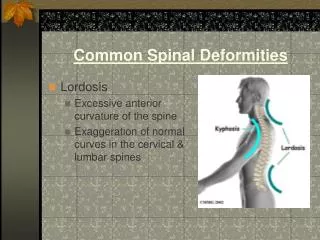
SpinalDeformities • Spinal deformities occur in either coronal or sagittal plane
What is scoliosis? • Lateral curvature of the spine >10º accompanied by vertebral rotation • Idiopathic scoliosis - Multigene dominant condition with variable phenotypic expression & no clear cause • Multiple causes exist for secondary scoliosis
Scoliosis classification 1-Structural: • Idiopatic • Congenital • neuromuscular • Neurofibromatosis • Osteochondrodystrophyos • Metabolic 2-Non-structural: (postural, histeric, herniopaty)
Idiopathic Scoliosis “Classification” • Age at Onset: Infantile: age birth to 3 years Juvenile: age 4 to 10 years Adolescent: age 11 to 17 years Adult: age 18 years up
Idiopathic Scoliosis “Etiology” • Remains unknown • Several studies have attempted to look into this and various factors have been postulated: genetic, tissue deficiencies, vertebral growth abnormalities, and central nervous system theories
Idiopathic Scoliosis “Genetic Factors” • Risenborough found a 11.1% incidence of scoliosis in first born relatives of patients with idiopathic scoliosis • Twins show a concordance of scoliosis with an incidence of 92% monozygotic and 63% dizygotic
Secondary causes for scoliosis:Inherited connective tissue disorders - Ehler’s Danlos syndrome - Marfan syndrome - Homocystinuria
Secondary causes for scoliosis:Neurologic disorders • Tethered cord syndrome • Syringomyelia • Spinal tumor • Neurofibromatosis • Muscular dystrophy • Cerebral palsy • Polio • Friedeich’s ataxia • Familial dysautonomia • Werdnig-Hoffman disease
Secondary causes for scoliosis:Musculoskeletal disorders • Leg length discrepancy • Developmental hip dysplasia • Osteogenesis imperfecta • Klippel-Feil syndrome
Characteristics of idiopathic scoliosis: • Present in 2 - 4% of kids aged 10 – 16 years • Ratio of girls to boys with small curves (<10º) is equal, but for curves >30º the ratio is 10:1 • Scoliosis tends to progress more often in girls (so girls with scoliosis are more likely to require treatment) • Toracal curve (right) • Lomber curve (left)
Natural history of scoliosis • Of adolescents diagnosed with scoliosis, only 10% have curve progression requiring medical intervention • Three main determinants of curve progression are: (1) Patient gender (2) Future growth potential (3) Curve magnitude at time of diagnosis
Natural history of scoliosis Assessing future growth potential using Tanner staging: Tanner stages 2-3 (just after onset of pubertal growth) are the stages of maximal scoliosis progression
Natural history of scoliosis Assessing growth potential using Risser grading: - Measures progress of bony fusion of iliac apophysis - Ranges from zero (no ossification) to 5 (complete bony fusion of the apophysis) - The lower the grade, the higher the potential for progression
Line Of Risser Risser 2 Risser 1 = 25% Capping. Risser 2 = 50% Capping. Risser 3 = 75% Capping. Risser 4 = 100% Capping. Risser 5 = 100% Capping + Fusion.
Natural history of scoliosis • Back pain not significantly higher in pts with scoliosis • Curves in untreated adolescents with curves < 30 º at time of bony maturity are unlikely to progress • Curves >50 º at maturity progress 1º per year • Up to 19% of females with curves >40 º have significant psychological illness • Life-threatening effects on pulmonary function do not occur until curve is >100 º (ie: Cor pulmonale)
Adam’s forward bend test • For this test, the patient is asked to lean forward with his or her feet together and bend 90 degrees at the waist. The examiner can then easily view from this angle any asymmetry of the trunk or any abnormal spinal curvatures .
Screening hints: • Shoulders are different heights – one shoulder blade is more prominent than the other • Head is not centered directly above the pelvis • Appearance of a raised, prominent hip • Rib cages are at different heights • Uneven waist • Changes in look or texture of skin overlying the spine (dimples, hairy patches, color changes) • Leaning of entire body to one side • Cavus feet
Red flags on PE: • Left-sided thoracic curvature • Pain • Significant stiffness • Abnormal neurologic findings • Stigmata of other clinical syndromes associated with curvature • Juvenilscoliosis MRI
Measure spinal curvature using Cobb method: • Choose the most tilted verterbrae above & below apex of the curve. - Angle b/t intersecting lines drawn perpendicular to the top of the superior vertebrae and bottom of the inferior vertebrae is the Cobb angle.
Treatment Decisions Accepted Standards
TLSO CTLSO
Brace Treatment for Scoliosis • Most common is Boston brace • Braces have 74% success rate at halting curve progression (while worn) • Bracing does not correct scoliosis, but may prevent serious progression • Usually worn until patient reaches Risser grade 4 or 5
Brace Treatment for Scoliosis • Of patients with 20 º - 29 º curves, only 40% of those wearing braces ultimately required surgery, compared to 68% of those not wearing back braces • Length of wearing time correlates with outcome (At least 16 hrs per day leads to best chance of preventing curve progression)
Idiopathic Scoliosis SURGICAL CORRECTION GOALS • Reduce the magnitude of the curve • Obtain fusion to prevent progression • Create a well-balanced spine
Idiopathic Scoliosis SURGICAL CORRECTION INDICATIONS • Curves over 45 degrees • Trunk deformity(rotation) • Trunk balance • Progressive curves despite bracing • Congenital scoliosis • Neurologic symptoms
Idiopathic Scoliosis GENERAL GUIDELINES FOR TREATMENT OF SCOLIOSIS • Under 20 degree’s: observe • 20 to 30 degree’s: observe with frequent follow-up; progression then brace • 30 to 45 degree’s: brace unless Risser 4/5 then observe • 45 plus degree’s: instrumentation
Surgical Treatment for Scoliosis • Curves in growing children greater than 40 º require a spinal fusion (Risser grade 0 to 1 in girls and Risser 2 or 3 in boys) • Skeletally mature patients can be observed until their curves reach 50 º • Posterior spinal fusion is best choice for thoracic curves • Anterior spinal fusion is best treatment for thoracolumbar and lumbar curves
Scoliosis • Adolescent idiopathic scoliosis • Structural scoliosis presenting at or about the onset of puberty and before maturity • 80 % of cases of idiopathic scoliosis • Mostly (90%) in girls • Predictors of progression very young age marked curvature Risser sign