Download
1 / 31
310 likes | 326 Vues
Learn about central nervous system infections, including acute meningitis, encephalitis, and brain abscesses. Discover the key etiological agents, examination methods, and approaches for different neuroinfections.

E N D
Institute for Microbiology, Medical Faculty of Masaryk University and St. Anna Faculty Hospital in Brno Miroslav Votava Agents of urinary tract infections Lecture for 3rd-year students 26th October, 2012
Importance of central nervous system infections – revision • CNS infections – relatively rare, but can have a very serious course • Incidence bacterial meningitis: 2/100.000/yearviral meningitis: 10/100.000/year • Lethality bacterial meningitis, non-treated: >70 % treated: ~10 %
Etiology ofacute meningitis – revision I Always distinguish purulent meningitis (nearlyalways of bacterial origin) from aseptic one (usually of viral origin) Anamnesis Clinical disease Laboratory – above all the examination of CSF cytology (appearance and number of cells) biochemistry (proteins and glucose) microbiology (microscopy, antigens, culture)
Cytology and biochemistry of CSF Etiology ofacute meningitis – revision II
Etiology of purulent meningitis by the age in % Etiology of acute meningitis – revision III
Etiology of purulent meningitis by the age in % Etiology of acute meningitis – revision IV
Importance of purulent meningitis according to etiology (lethality and sequelae) Etiology ofacute meningitis – revision V
The most common agents of asepticmeningitis: VIRUSES mumps virus (but CNS infection is clinically silent) enteroviruses: echoviruses (30 serotypes) coxsackieviruses (23 + 6 serotypes) tick-borne encephalitis virus (TBEV) rarely HSV and VZV and other neuroviruses rarely some bacteria leptospirae, borreliae, Mycobacterium tuberculosis Etiology ofacute meningitis – revision VI
Overview of Central-European neuroviruses – revision TBEV (tick-borne enc. v.) other arboviruses enteroviruses: polio LCMV coxsackie /morbilli v./ echo /EBV/ mumps v. /polyomaviruses JC & BK/ HSV, VZV, CMV /HIV/ rabies v. /prions/
Arboviruses in Central Europe – revision II Arboviruses isolated in Czech Republic, probably nonpathogenic for humans: Bunyaviridae: Lednice Sedlec Other Europeanpathogenic arboviruses, which may be imported: dengue v. (flavivirus, Greece) CCHFV (nairovirus, Ukraine, Bulgaria) Toscana v. (phlebovirus, Italy) Bhanja v. (bunyavirus, Slovakia) chikungunya v. (alphavirus, Italy)
Etiology of chronic meningitis– revision • Bacteria: Mycobacterium tuberculosis (meningitis basilaris) Treponema pallidum • Moulds and yeasts: aspergilli Cryptococcus neoformans
Etiology of encephalitis– revision Encephalitis – only acute, of viralorigin: • tick-borne encephalitis v. • HSV • enteroviruses • mumps v.
Etiology of acute brain abscess – revision Acute brain abscesses are only of bacterialorigin: • mixed anaerobic and aerobic flora • staphylococci (both S. aureus and coagulase negative staphylococci) • group A and D streptococci
Etiology of chronic brain abscess – revision bacteria: Mycobacterium tuberculosis Nocardia asteroides mycotic organisms: Cryptococcus neoformans (yeast) parasites: Cysticercus cellulosae (tissue form of pork tapeworm Taenia solium)
Microbiological examination of CNS infections – addendum I • PURULENT MENINGITIS Cerebrospinal fluid(CSF) microscopy: Gram; sometimes Ziehl-Neelsen; Heidenhein; Indian ink; wet mount detection of antigens: N. meningitidis A, C, Y, W135, B (Ag asE.coli K1); S. pneumoniae; GBS; H. influenzae b; Cryptococcus neoformans culture:blood agar (BA)with staph. line; chocolate agarin 5% CO2; BA anaerobically; Endo or MacConkey; if requiredmediafor mycobacteria, cryptococci, amoebae etc. Blood for haemoculture notably positive in meningococcal meningitis
Microbiological examination of CNS infections – addendum II 2. OTHER NEUROINFECTIONS Viral neuroinfections (asept. meningitis & enceph.): CSF for the direct virus detection (PCR, tissue culture) blood for the antibody detection (IgM, 4fold titre rise): in susp. infections with TBEV, HSV 1 and 2, VZV, CMV, mumps virus Borreliosis and neurosyphilis: blood for the antibody detection Brain abscess (both acute and chronic): punctateorexcissionfor bacteriology (incl. tbc), mycology (cryptococci) & histology (cysticerci)
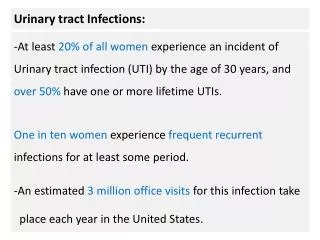
Urinary tract infections (UTIs) Frequency of UTIs: The 2nd most common infections (after respiratory ones) In adults: the most commoninfections in a general practitioner’s office Afflicting mainly females(because of shorter urethra)
Examples of UTIs The most common UTI: cystitis develops ascendently caused by intestinal microflora main symptoms: dysuria (difficult urination with sharp and burning pain) pollakisuria (urgent need to urinate accompanied by urination of a small amount of urine only) Other UTIs: mainly pyelonephritis(more serious) origin: ascendent or hematogenous urethritis – will be dealt with as STD
Etiology of UTIs Proportional representation of microbesdiffers in • non-complicated UTIs • infections accompanying structuralabnormalities (prostatic hypertrophia, urinary stones, strictures, pregnancy, congenital defects, permanent catheters) • infections accompanying functionaldisorders (vesicoureteral reflux, neurological disorders, diabetes mellitus)
Etiology of non-complicated UTIs circa 80 % Escherichia coli circa 10 % enterococci (Enterococcus faecalis) circa 5 % Proteus mirabilis rest: other enterobacteriae (Klebsiella pneumoniae, Kl. oxytoca, Ent. cloacae, C. freundii etc.) Streptococcus agalactiae coagulase neg. staphylococci (S. epidermidis, S. saprophyticus, S. haemolyticus etc.) yeasts (mainly Candida albicans)
Etiology of complicated UTIs circa 80 %: Escherichia coli Klebsiella pneumoniae Proteus mirabilis Pseudomonas aeruginosa enterococci the rest: other enterobacteriae acinetobacters other G-neg. non-fermenting rods candidae
Lege artistaking a urine sample • Only after a thorough cleaning of genital, incl. external orificium of urethra by means of soap and water • Take the middle stream of urine only • Use a sterilevessel • Pour urine into a steriletube& stopper it promptly • If not possible to process it within 2 hours, place the specimen into 4 °C for 18 hours at most
Semi-quantitative examination of the urine sample – I We are interested • not only in the kind of microbe present in the urine sample, but especially in • the amount of the microbe Why are we interested in the number of microbes in 1 ml of urine? Because • high numbers only stand for the UTI • low numbers mean usually contamination acquired during urination
Semi-quantitative examination of the urine sample – II Therefore, the urine is inoculated on culture media by means ofcalibrated loop, usually taking exactly 1 μl of urine In this case 1 colony means 103 CFU/ml 10 colonies mean 104 CFU/ml 100 colonies mean 105 CFU/ml (CFU = colony-forming unit = 1 bacterial/yeast cell)
Media used in microbiological examination of urine • Blood agar inoculated by means of calibrated loop • Chromogenic medium oriented on the most frequent urinary pathogens; their colonies are of different colour inoculated by means of calibrated loop • According to requirements further media e.g. chromogenic medium for yeasts or a medium for MRSA
Primary urine pathogens Escherichia coli & most of other enteric bacteria enterococci (mostly Enterococcus faecalis) Streptococcus agalactiae staphylococci (mostly coagulase negative: S. epidermidis, S. saprophyticus, S. haemolyticus etc.; but also S. aureus) yeasts (in the main Candida albicans) Pseudomonas aeruginosa & some other Gram-negative non-fermenting rods . . .
Homework 5 – solution Jacques-Louis David (1748-1825): Death of Marat (1783) What is the connection between this painting and medicine? • Jacques-Louis David, had a facial tumor • Jean Paul Marat, murdered by Charlotte Corday in 1793, was initially a physician • He was run through when taking a bath for treatment his skin disorder (probably dermatitis herpetiformis Dühring)
Homework 6Who is the author of this painting and what is its name?
Answer and questions The solution of the homework and possible questions please mail to the address mvotava@med.muni.cz Thank you for your attention