Download
1 / 26
260 likes | 310 Vues
Learn about respiratory viruses, bacterial agents, and etiology of respiratory illnesses in a comprehensive lecture for dentistry students. Understand the causes of bronchitis, bronchiolitis, pneumonia, and more.

E N D
Institute for Microbiology, Medical Faculty of Masaryk University and St. Anna Faculty Hospital in Brno Miroslav Votava Agents of digestive system infections – I The 3rd lecture for 3rd-year students of dentistry 6thDecember, 2010
Respiratory viruses – revision • The most important and most common: • influenzavirus A a B • adenoviruses • RSV and metapneumoviruses • parainfluenzaviruses (type 1+3 = Respirovirus, type 2+4 = Rubulavirus) • rhinoviruses • coronaviruses (incl. SARS agent)
Other respiratory agents of virological interest – revision • Bacterial agents causing atypicalpneumoniae (but diagnosed in virological laboratories): • Mycoplasmapneumoniae– the most common • Coxiellaburnetii – Q-fever • Chlamydiapsittaci – ornithosis • Chlamydiapneumoniae
Etiology of epiglottitis – revision • Epiglottitis acuta: Serious disease – medical emergency The child may suffocate! • Practically one and only important agent: Haemophilus influenzae type b
Etiology of laryngitis and tracheitis – revision • Again respiratoryviruses but other than agents of nasopharyngitis: parainfluenza and influenzaA viruses & RSV • Bacteria: C. pneumoniae, possibly Mycopl. pneumoniae, secondarily: S. aureus and Haem. influenzae laryngotracheitis pseudomembranosa (croup): Corynebacterium diphtheriae
Etiology of bronchitis – revision • Acute bronchitis: Viruses:influenza, parainfluenza, adenoviruses, RSV Bacteria, secondarily after viruses: pneumococci, Haem. influenzae, Staph. aureus, Moraxella catarrhalis Bacteria, primarily: Mycoplasmapneumoniae, Chlamydia pneumoniae, Bordetellapertussis • Chronic bronchitis (cystic fibrosis): • Pseudomonas aeruginosa, Burholderia cepacia
Etiology of bronchiolitis – revision • Isolated bronchiolitis in newborns and infants only: Pneumovirus (= respiratory syncytial virus = RSV) Metapneumovirus
Different types of pneumoniaehave different etiologies • Acute – community-acquired pneumoniae • in originally healthy (1) • adults (2) • children (3) • in debilitated persons (4) • after a contact with animals (5) • Acute – nosocomialpneumoniae – VAP = ventilator-associated (6) • early (7) • late (8) –others (9) 3. Subacute and chronic pneumoniae(10)
Etiology of pneumoniae I– revision • Acute,community-acquired, in healthy adults • bronchopneumonia and lobar pneumonia: • Streptococcus pneumoniae • Staphylococcus aureus • Haemophilus influenzae type b • atypicalpneumonia: • Mycoplasma pneumoniae • Chlamydia pneumoniae • Influenza A virus (during an epidemic only)
Etiology of pneumoniae II– revision • Acute, community-acquired, in healthy children • Bronchopneumonia: • Haemophilus influenzae • Streptococcus pneumoniae • Moraxellacatarrhalis • In newborns: Streptococcus agalactiae enterobacteriae • atypical pneumonia: • respiratory viruses (RSV, infl. A, adenoviruses) • Mycoplasma pneumoniae • Chlamydia pneumoniae • in newborns: Chlamydia trachomatis D-K
Etiology of pneumoniae III – revision • Acute, community-acquired, in debilitated individuals: • pneumococci, staphylococci, haemophili • Klebsiellapneumoniae (alcoholics) • Legionellapneumophila • In more serious immunodeficiency: • Pneumocystis jirovecii • CMV • atypical mycobacteria • Nocardia asteroides • aspergilli, candidae
Etiology of pneumoniae IV – revision • Acute, community-acquired, after a contact with animals: • Bronchopneumonia • Pasteurella multocida • Francisella tularensis (tularemia) • Atypical pneumonia • Chlamydia psittaci (ornithosis) • Coxiella burnetii (Q-fever)
Etiology of pneumoniae V– revision • Acute, nosocomial: • VAP (ventilator-associated pneumonia) • early (up to the 4th day of hospitalization): sensitive communitystrains of respiratory agents • late (from the 5th day of hospitalization): resistant hospital strains • Others • viruses (RSV, CMV) • legionellae
Etiology of pneumoniae VI– revision • Subacute and chronic: • aspiration pneumonia and lung abscesses • Prevotella melaninogenica • Bacteroides fragilis • peptococci and peptostreptococci • lung tuberculosis and mycobacterioses • Mycobacterium tuberculosis • Mycobacterium bovis • atypical mycobacteria(e.g. the complex M. avium–M. intracellulare) …
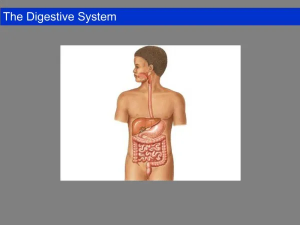
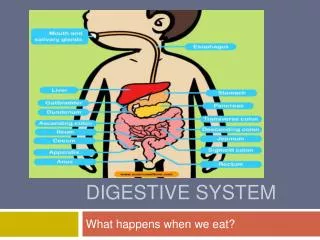
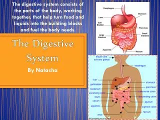
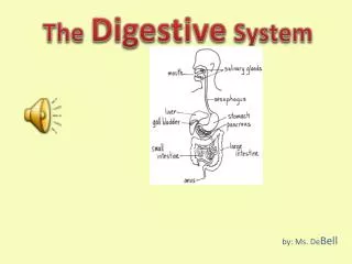
Digestivesystem • a microbiologist´s dreamland • a fruitful microbial garden • its both ends are the „buggiest“ parts of the body • in the colon: approx. 1012 bacteria/g • normal colonic flora: 99 % anaerobes(Bacteroides, Fusobacterium, Clostridium, Peptostreptococcus), only 1 % enteric bacteria (mostly E. coli)& enterococci
Mouth cavity – I Normal flora: • viridans (= α-haemolytic) streptococci (e.g. Streptococcus salivarius) • oral neisseriae (e.g. Neisseria subflava) • haemophili of very low pathogenicity (e.g. Haemophilus parainfluenzae) Dental plaque: adherent microbial layer at the tooth surface consistingof living and dead bacteria and their products together with components from the saliva In essence, dental plaque is a biofilm It cannot be washed off, only mechanically removed
Mouth cavity – II Dental caries: chronic infection caused by normal oral flora → localized destruction of tooth tissue Etiology: mouth microbes (mostly Strept.mutans) making acids from sucrose in food Thrush (in Latin soor): Candida albicansItoccursmostly in newborns Herpetic stomatitis: primary infection with HSV 1 Ludwig´s angina: polymicrobial anaerobic infection of sublingual and submandibular spaces (Porphyromonas, Prevotella etc.)
Oesophagus Infections never occur in previously healthy individuals Only in severely immunocompromised persons (AIDS or after chemotherapy): • Candida albicans • Cytomegalovirus (CMV)
Stomach Stomach = a sterilization chamber killing most of the swallowed microbes by means of HCl Exception: Helicobacter pylori It produces a potent urease and by splitting tissue urea it increases pH around itself (1 molecule of urea → 1 CO2 + 2 NH3) H. pylori causes • chronic gastritis • peptic ulcers (Warren & Marshall, Nobel price in 2005)
Biliary tree & the liver – I Acute cholecystitis (colic, jaundice, fever): obstruction due to gallstones Etiology: intestinal bacteria (E. colietc.) Complication: ascending cholangitis Chronic cholecystitis: the most dangerousagent is Salmonella Typhi (carriers of typhoid fever) Granulomatous hepatitis: Q fever, tbc, brucellosis
Biliary tree & the liver – II Parasitic infections of the liver: Amoebiasis (Entamoeba histolytica: liver abscess) Malaria (the very first, clinically silent part of the life cycle of malaric plasmodia) Leishmaniasis (Leishmania donovani:kala-azar, L. infantum) Schistosomiasis (eggs of Schistosoma japonicum, less often S. mansoni)
Systemic infections which start in the digestive tract Enteric fever (typhoid fever and paratyphoid fever): Salmonella Typhi, Salmonella Paratyphi A, B and C Listeriosis: Listeria monocytogenes Peritonitis: colonic flora (Bacteroides fragilis + other anaerobes + mixture of facultative anaerobes) Viral hepatitis: HAV, HBV, HCV, HDV, HEV
Small and large intestine Bacterial overgrowth syndrome: After surgery, during depressed peristalsis or gastric achlorhydria bacteria may overgrow in the small intestine → steatorrhea, deficiency of vitamin B12, diarrhea, malabsorption of vitamins A and D Diarrhea: increase in daily amount of stool water – common intestinal response to many agents Dysentery: acute inflammation of the colon → abdominal pain & small-volume stools with blood, pus and mucus
Etiology of diarrheal disease Infectious etiology: • Bacterial (most frequent) • Viral • Parasitic • Mycotic Non-infectious etiology: • Food poisoning . . .
Answer and questions The solution of the homework and possible questions please mail (on 6.30 a.m. at the latest) to the address mvotava@med.muni.cz Thank you for your attention