Download
1 / 60
600 likes | 620 Vues
Learn about the complex pathophysiological process of concussion and its causes, symptoms, evaluation, imaging, and management. Discover risk factors, grading systems, and guidelines for return to play.

E N D
Concussionand Neurologic Injury Jamie B. Varney, M.D. CAQ Sports Medicine Pikeville Medical Center Orthopedics and Sports Medicine
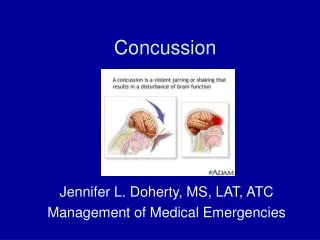
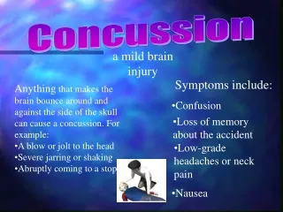
What is a Concussion? • Complex pathophysiological process affecting the brain, induced by traumatic biomechanical forces1
Cause of Concussion • May be caused by direct blow to head, face, neck or elsewhere • Thought to be due to axonal injury caused by acceleration forces • Not typically a structural injury • Electrolyte shifts and release of neurotransmitters and free radicals thought to play role • Fuel need/delivery mismatch
Risk Factors • Previous concussion (strongest factor) • Improper technique • Male > Female
High Risk Sports • Football • Ice Hockey • Soccer • Boxing • Rugby • Field Hockey • Lacrosse
Symptoms • Headache • Loss of consciousness • Confusion/Memory Loss • Dizziness/Vertigo • Nausea/Vomiting • Phono/photo phobia • Incoordination/Slowed reaction • Emotional lability/irritability • Sleep disturbance
Symptoms Confusion • Vacant stare • Slow response • Easily distracted • Decreased focus • Disoriented • Slurred speech
Symptoms • Memory Deficits • Repeats questions • Retrograde amnesia • Anterograde amnesia (inability to form new memories)
Rare Symptoms • Seizure 1% or less • Cortical blindness
Evaluation • Should be evaluated by trained personnel as soon as suspected injury • On Field • Loss of Consciousness • ABC’s • Rule out C-Spine injury • assumed if LOC • Neurological Status • Mental Status
Mental Status • Orientation • Memory • Cognitive skills
Memory • Short term • Events of game (plays/score) • Word recall • Number sequence recall • Intermediate • Delayed word recall • Previous games • World events • Long term • Teammates/Family members • Birthdates • Presidents
Cognitive skills • Serial 7’s • Reverse spelling • Reverse alphabet • Concentration / complex commands
Neurological function • Cranial Nerves • Motor • Sensory • Reflexes • Cerebellar function/Coordination • Finger/nose • Heel/shin • Gait/Tandem (eyes closed as well) • Rhomberg/ Pronator drift
Additional Exam • Skull for depressions • Cervical spine tenderness • Nose for clear drainage • Ears for hemotympanum • Signs of skull fracture
Sideline Tools • SCAT3>13 y/o • Standardized Assessment of Concussion (SAC) • Maddock's Questions • Modified BESS • Balance Error Scoring System • Child SCAT3 <13 y/o
Neuroimaging • Typically normal • CT preferred if necessary • MRI more sensitive but may not correlate with severity or outcome • Possible future role for functional MRI
Recommended Imaging • Neurological deficit • Suspected C-Spine injury • Suspected skull fracture • Raccoon eye’s • Battle’s Sign • Rhinorhea • Hemotympanum • Seizure • Coagulopathy / Anticoagulant use • Progressive symptoms
Consider Imaging • Canadian CT criteria • GCS <15 two hours after injury • Two or more episodes vomiting • Age > 65 • Amnesia longer than 30 min prior • Dangerous mechanism • MVA • Fall > 3ft or 5 stairs
Consider Imaging • New Orleans Criteria (GCS 15) • Headache • Vomiting • Age >60 • Drug/ETOH intoxication • Persistent anterograde amnesia • Visible trauma above clavicle
Comparison • Two studies have shown both are 100% sensitive for detecting neurosurgical abnormalities • One study showed higher sensitivity for clinically significant findings with New Orleans (99.4% vs 87.2%) • Canadian CT rules more specific • Lowered CT rates 52.1% versus 88% • Other study specificity 39.7% vs 3%
Bottom Line1 Imaging usually not helpful for concussion Helpful to rule out bleeds if progressive symptoms or clinical suspicion
Hospital Admission • GCS <15 • Abnormal CT scan • Seizures • Bleeding diasthesis or anticoagulants • Consider if no one available to monitor for progression of symptoms
Outpatient Monitoring • Monitor Closely 1st 24 hrs • Educate about warning signs • Somnolence/Confusion • Worsening headache • Vision difficulties • Vomiting or stiff neck • Neurological deficits • Avoid strenuous activity
Grading Concussion • Old system • Colorado • American Academy of Neurology (AAN) • Cantu • Prague Statement 2004 • Simple <10 days • Complex >10 days/seizures/prolonged LOC • Zurich Statement 2012 • Forget Grades
Return to Play1 • No same day play • KHSAA and NCAA • Physical Rest Until Asymptomatic • Consider Cognitive Rest • Exercise Testing
Progressive Return To Play1 • Step 1 • No activity, rest, when symptom free without meds go to step 2 • Step 2 • Light aerobic exercise, no resistance training • Step 3 • Sport specific exercise • Step 4 • Non Contact Practice and Resistance Training • Step 5 • Full Contact Practice • Step 6 • Full Game
Office Exertional Maneuvers • Treadmill/Bike • Sprints/Run in place • Sit-ups, Push-ups
Progressive RTP • If symptoms develop at any step stop and rest. Do not proceed. • ATC's are invaluable resource • More conservative in children with focus on cognitive rest and return to learn before return to play
Second Impact Syndrome • Occurs after second injury before first injury has healed • Diffuse cerebral swelling that can be life threatening • Few cases with documentation that is consistent with description • May only require minor injury
Post traumatic Epilepsy • Seizure within 1st week not epilepsy • Mild TBI associated with twofold risk epilepsy in 5 years
Post Concussive Syndrome • Not related to severity of injury • Symptoms >3 months (DSMIV) • Headache • Dizziness • Fatigue • Irritability • Anxiety/Depression • Insomnia • Loss of concentration or memory • Cognitive impairment
Post Concussive Syndrome • Treatment • Consider referral • Treat symptoms
Mood Disorders Dementia Movement Disorders Chronic Traumatic Encephalopathy (CTE)
Neuropsychiatric Testing • Paper tests interpreted by experienced neuropsychologist • Computerized Tests
Neuropsychiatric Testing • Speed of information processing • Memory • Attention • Concentration • Reaction Time • Scanning • Visual tracking • Problem solving
Neuropsychiatric Testing • Tested at baseline then post injury if needed • More sensitive than classic testing • Concern is maybe too sensitive and not specific enough
Prevention • Proper equipment / fitting • Proper training for coaches and support staff • Enhancement and enforcement of protective rules • Pre-participation evaluation of concussion history
Other Neurological Injury C-Spine Brachial Plexus Transient Cord Neuropraxia
Other Neurological Injury C-Spine Brachial Plexus Transient Cord Neuropraxia
Brachial Plexus Injury • Commonly called stinger / burner • Caused by stretch or compression • Unilateral symptoms • Weakness • Numbness • Stinging pain • C5-6 most common • If has bilateral symptoms think cord injury
Brachial Plexus Injury • Single episode • May return when no pain or neurologic deficit • Recurrent episode • Consider evaluation including flex/ext x-rays and canal diameter • If symptoms last more than 1 week consider MRI/EMG to rule out cord lesion
Prevention • Rehab to strengthen neck/shoulders • Proper hitting technique • Proper equipment (pads) • Neck rolls/cowboy collars
Transient Cord Neuropraxia • Flexion/extension injury with underlying spinal stenosis • Post traumatic neurological findings • Bilateral symptoms of paresthesia and or weakness • Upper > Lower extremities • Lasts minutes to days • If occurs must evaluate with imaging for cord injury and spinal canal diameter
Torg Ratio • Ratio of spinal canal to vertebral body • Ratio <0.8 suggestive of stenosis • MRI measurement of cord vs. canal diameter more reliable
Treatment • If have transient neuropraxia then protect cervical spine until fracture ruled out • Must evaluate canal diameter which may imply risk of future injury • Neurosurgeon familiar with treatment should help make any return to play decision
References • McCrory,P. et al. Consensus Statement on Concussion in Sport (Zurich Statement 2012). Br J Sports Med 2013;47:250-258 • Meehan, WP, O'Brien, MJ. Sports-Related Concussion in Children and Adolescents: Clinical Manifestations and Diagnosis. UpToDate. 9-22-14