Download
1 / 91
960 likes | 1.3k Vues
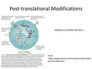
Post-translational modifications PC235 Katalin F. Medzihradszky folkl@cgl.ucsf.edu. Post-translational Modification of Proteins Expanding Nature’s Inventory (2006) by C.T. Walsh ISBN 0-9747077-3-2. Just for introduction. Genome ↓ Transcription – mRNA ↓

E N D
Post-translational modificationsPC235Katalin F. Medzihradszkyfolkl@cgl.ucsf.edu Post-translational Modification of Proteins Expanding Nature’s Inventory (2006) by C.T. Walsh ISBN 0-9747077-3-2
Just for introduction Genome ↓ Transcription – mRNA ↓ Translation – proteins + co-translational modifications ↓ Post-translational modifications
Post-translational modifications I • Enzymatic processing • N-, O-, C-linked glycosylation – Asn, Ser/Thr/Hyl/Hyp, Trp • Phosphorylation – Tyr, Ser, Thr, His, Asp • Acylation acetylation of the N-terminus fatty acid anchors on Cys • Cross-linkage – Lys, Trp, Tyr, Met • Oxydation – Cys, Met, Trp, Tyr, His • Methylation – N-terminus, Arg, Lys • Ubiquitination - Lys
Post-translational modifications II • Cannot be predicted – though consensus sequences have been reported for some of them • Organism-dependent • Can be tissue- or location-specific • Stable or dynamic – high and low level • Alters biological activity, and physical properties • May alter the immune response ↓
Sample preparation • Will the modification survive? • Can I get it down to a mass spectrometry friendly size? • Isolation/enrichment? • Losses – too hydrophilic/hydrophobic
Mass spectrometry • Will the modification survive the ionization? the MS/MS activation? • How unambiguous is the assignment? modification assignment? Ac vs. Me3; phosphate vs. sulfate etc. site assignment?
Finding the N-termini • 80-90% of the eukaryotic proteins is acetylated If 2nd aa: G, A, S, C, T, P, V • Met-1 is clipped off; Ac- is added to G, A, S, T If 2nd aa: E, D, Q, M, I, L, W, F – persisting Met gets acetylated
Finding the N-termini • McDonald L, et al., Positional proteomics: selective recovery and analysis of N-terminal proteolytic peptides.Nat Methods. 2005 Dec;2(12):955-7. Epub 2005 Nov 18.
Modified N-terminus Modified side-chain I don’t think any search engine will tell the difference !
Methylation (mono, di, tri - +14, +28, +42 Da) • N-terminus, Lys, Arg, His • Trimethyl – acetyl = 36 mmu Accurate mass measurement helps; Fragmentation is different too • Glu (Asp) may form Me-ester – upon CBB staining (MeOH + acid) + 14 Da
Disulfide-bridges • in membrane and secreted proteins important 3D structure feature • prone to shuffling @ basic pH
Assigning disulfide-bridges I Reduction/alkylation Reduction/alkylation m/z I Digestion @ low pH Digestion @ low pH m/z I Reduction/alkylation MS/MS I Reduction/alkylation m/z I m/z m/z
Synthetic Ac-TIMP-1(Ser175)126-184 ECTVFPCLSIPCKLQSGTHCLWTDQLLQGSEKGFQSRHLACLPREPGLCS WQSLRSQIA Where are the disulfide bridges? * digestion with trypsin @ pH 6; * with pepsin in acid Bodi, N. et al., J. Pept. Sci.9, 430-441 (2003).
[33-37] 41-49 [38-44]-[45-55] intensity [38-44] Ac[1-13]-[45-55] 2-7, 12-49 20-41 [45-55] [14-32]-[38-44] free SH 41-49 [38-55] Free SH 600 800 1000 1200 2000 2200 2400 2600 2800 3000 m/z m/z S* [45-55] 2 S* [38-55] 3 S* Ac[1-13] intensity 1 S* [14-37] 1 S* [14-32] Ac[1-13] 1 S-S, 1 S* Ac[1-13] 1free SH, 2 S* 2 S* S* [38-59] [38-44] 1 S* [33-37] [45-59] 1600 1800 2000 2200 2400 2600 2800 3000 600 800 1000 1200 1400 1600 m/z m/z MALDI-TOF analysis of the tryptic digest In-source reduction PSD
Results from the MALDI-TOF MS Tryptic digest After reduction/alkylation
Disulfide bridges in TIMP-1 C-terminal domain: [38-44]-S-S-[45-55], PSD of MH+ at m/z 2082.1 MALDI-PSD/CID yields characteristic triplets
Disulfide Bridges The ProteinProspector mass modification search can be used in conjunction with MS-Bridge to find peptides with disulfide bridges. For this example, mass shifts between 0-2000 Da were considered. (1023.3276+4). KLSWADLIVFAGNCALESMGFK+4 VSFADLVVLGGCAAIEK
Glycosylationhttp://glycores.ncifcrf.gov/Reference: Essentials of Glycobiologyby Varki et al.
N-linked AsnXxxSer/Thr/Cys
N-linked glycosylation • consensus sequence • GlcNAc2Man3 – core oligomannose structure – just Man units complex sugars– GlcNAc-Gal–SA antennae hybrid structures core fucosylation sulfate, phosphate modifications • PNGase F removes all N-linked structures; Asn Asp
N-linked glycosylation • Incredible heterogeneity: a site may be only partially occupied and may display numerous different carbohydrates • species-, tissue-, cell-type-specificmodification, physiological changes, diseasesmay alter the sugars
certain structures are immunogenic Gal a1-3 capping, Fuc a1-3 on inner GlcNAc; blood group determinants
N-linked glycosylation • Identification from diagnostic fragments: * HexNAc m/z 204 * HexHexNAc m/z 366 Precursor scan, or „ping-pong” acquisition • Identification from oligosaccharide heterogeneity • enrichment by HILIC or lectin-chromatography
human lecithin:cholesterol acyltranferase and apolipoprotein D, tryptic digest, LC/MS analysis
Recombinant Factor VIII, 50 kDa subunit MHTVNGYVN*R
AG(Man8GlcNAc2)NVSNIIPASATLNADVR peptide+GlcNAc
About the structures of N-linked glycopeptides • from the measured mass, and the CID spectrum the modified peptide can be identified + the size and class of the sugar • the identity of the sugar units and their linkage positions CANNOT be determined • NMR, exo- and endoglycosidases are needed to complete the job
[MHNa2]3+ of QV(Man10GlcNAc2)NIT and [MH2Na] 3+ of QV(Man9GlcNAc2)NITGK
One component from the previous slide K.F. Medzihradszky Meth. Enzymol.405, 116-138 (2005).
O-linked sugars • No consensus sequence • No common core structure • No universal enzyme b-elimination works (NaOH) sugars have to be reduced upon release Detection is problematic – because of heterogeneity; variable site occupancy Site assignment is even harder
Other O-linked core structures • Fuc Harris, R.J. & Spelmann, M.W. (1993) Glycobiology,3, 219-224. • Glc Nishimura, H et al., (1989) J. Biol. Chem.264, 20320-20325. • Man – in yeast ____________________________________ • GlcNAc – single unit; INSIDE the cell
J. Am. Soc. Mass Spectrom. 7, 1996, 319-328. CID fragmentation of O-linked glycopeptides
GlcNAc GlcNAc protein GlcNAc O-GlcNAcylation O-GlcNAc PSVPVSerGSAPGR WGA O-linked GlcNAc • Regulatory modification of nuclear and cytoplasmic proteins • Poorly understood due to lack of effective methods for enrichment and detection. • The Enrichment Problem • WGA lectin has affinity for GlcNAc, but affinity to a single GlcNAc moiety is low: millimolar1. Complex glycosylation Good recovery of glycoprotein Load Wash Elute WGA No recovery Load Wash Elute Ohlson S et al. Bioseparation (1998) 7 p.101
UV absorbance 214 O-GlcNAc modified enriched Non-enriched Time (minutes isocratic HPLC) O-GlcNAc Enrichment • Selective Enrichment of O-GlcNAc modified peptides using lectin weak affinity chromatography1. Peptide Mixture Isocratic Chromatography fraction collection WGA column Vosseller, K. et al. Mol Cell Proteomics (2006) 5 5: p.923-934
CID Analysis of O-GlcNAc-Modified Peptides • O-glycosidic link is significantly more labile under CID conditions than peptide backbone. • Modification site identification using CID often not possible. Chalkley, R. J. and Burlingame, A. L. J. Am. Soc. Mass Spectrom. (2001) 12 p.1106-1113
A bit about MS/MS alternatives • ECD (electron-capture dissociation) – multiply charged ions meet electron beamin FT-ICR – larger the charge state larger the capture’s efficiency • ETD (electron-transfer dissociation) – multiply charged ions meet stable anion (fluoranthene) in ion traps • radical ion is formed, different mechanism mostly backbone cleavages
Bakken, V.; Helgaker, T.; Uggerud, E., Eur. J. Mass Spectrom. 2004, 10, 625-638 Mechanism of ECD II
ECD MS Spectrum of GlcNAc-modified Peptide from Spectrin Vosseller, K et al. Mol Cell Proteomics (2006) 5 5: p.923-924
PhosphorylationBiological significance • One of the most important regulatory events: • turns proteins on and off • induces or prevents other post-translational modifications in the same protein • signaling pathways : phosphorylation cascades
Difficulties • Dynamic process : kinase vs. phosphatase • – both must be blocked during isolation • Phosphorylation often @ low level (<5%) • Lower ionization efficiency – signal of phosphopeptides suppressed Enrichment is a must at protein level at peptide level
A whole cell lysate with 20,000 sites of phosphorylation at 1% stoichiometry
A whole cell lysate with 20,000 sites of phosphorylation at 1% stoichiometry
phosphopeptide relative distribution after a 10,000 fold enrichment