Post-translational modifications
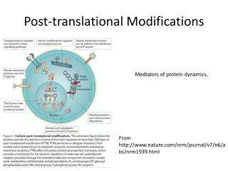
Post-translational modifications. Post-translational Modification of Proteins Expanding Nature’s Inventory (2006) by C.T. Walsh ISBN 0-9747077-3-2. Just for introduction. Genome ↓ Transcription – mRNA ↓ Translation – proteins + co-translational modifications ↓

Post-translational modifications
E N D
Presentation Transcript
Post-translational modifications Post-translational Modification of Proteins Expanding Nature’s Inventory (2006) by C.T. Walsh ISBN 0-9747077-3-2
Just for introduction Genome ↓ Transcription – mRNA ↓ Translation – proteins + co-translational modifications ↓ Post-translational modifications
Post-translational modifications I • Enzymatic processing • N-, O-, C-linked glycosylation – Asn, Ser/Thr/Hyl/Hyp, Trp • Phosphorylation – Tyr, Ser, Thr, His, Asp • Acylation acetylation of the N-terminus fatty acid anchors on Cys • Cross-linkage – Lys, Trp, Tyr, Met • Oxydation – Cys, Met, Trp, Tyr, His • Methylation – N-terminus, Arg, Lys • Ubiquitination - Lys
Post-translational modifications II • Cannot be predicted – though consensus sequences have been reported for some of them • Organism-dependent • Can be tissue- or location-specific • Stable or dynamic – high and low level • Alters biological activity, and physical properties • May alter the immune response ↓
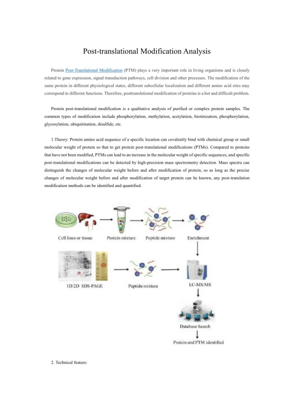
How to find PTMs? Sometimes proteins give us a clue: • higher/lower/wider band on SDS-PAGE For example, +~5 kDa on SDS-PAGE may indicate 1 occupied N-linked glycosylation site • multiple spots with IEF or on 2D-gel • higher MW with gel-filtration or native gel-elfo • altered hydrophobicity • altered immunochemistry • altered biological activity • “blank” cycle in Edman sequencing (with proper standard & altered chromatography the modification could be identified: Identification of glycosylation sites in mucin peptides by edman degradation. Zachara NE, Gooley AA. Methods Mol Biol. 2000;125:121-8. Review.
How to find PTMs? Sometimes we’re searching for something specific • using a known donor and radioactive labeling ATP for phosphorylation (Why cannot we use this approach for glycosylation?) • using modification-specific derivatization and staining or enrichment Periodate oxidation and labeling for glycosylation • using modification-specific antibodies Against phosphoTyr • using modification-specific enzymes Phosphatase or glycosidase treatment • Mass spectrometryMass Spectrometry Mass Spectrometry!
Sample preparation • Does the modification survive? • Losses due to enzymatic reactions Phosphatase activity for phosphopeptides • Losses due to chemical reactions Acid-sensitive modifications, such as phosphoHis • Losses due to different physical properties Fatty-acylated or glycopeptides • Do we introduced some modifications? * Biological artifacts * Chemical side reactions
Some modifications that occur in vivo and can be introduced in vitro • Carbamidomethylation of N-termini and Lys side-chains • Formylation of Lys side-chains • Asn cyclization; b-aspartame formation; deamidation • Oxidation • Glu, Asp methyl ester formation And the list goes on…
Finding the N-termini • 80-90% of the eukaryotic proteins is acetylated If 2nd aa: G, A, S, C, T, P, V • Met-1 is clipped of; Ac- is added to G, A, S, T If 2nd aa: E, D, Q, M, I, L, W, F – persisting Met gets acetylated
Finding the N-termini • N-terminal sequencing • Positive enrichment (Wells-group); subtiligase-mediated selective N-terminal labeling with biotin-containing probe; enrichment; release with TEV protease; MS-analysis “Global sequencing of proteolytic cleavage sites in apoptosis by specific labeling of protein N termini.” Mahrus S, Trinidad JC, Barkan DT, Sali A, Burlingame AL, Wells JA. Cell. 2008 Sep 5;134(5):866-76. • Negative enrichment – next slide
Finding the N-termini • McDonald L, et al., Positional proteomics: selective recovery and analysis of N-terminal proteolytic peptides.Nat Methods. 2005 Dec;2(12):955-7. Epub 2005 Nov 18.
Modified N-terminus Modified side-chain
Methylation (mono, di, tri - +14, +28, +42 Da) • N-terminus, Lys, Arg, His • Trimethyl – acetyl = 36 mmu Accurate mass measurement helps; Fragmentation is different too • Glu (Asp) may form Me-ester – upon CBB staining (MeOH + acid) + 14 Da
Disulfide-bridges • in membrane and secreted proteins important 3D structure feature • Some toxins and antibacterial peptides feature characteristic Cys network • -S-S- prone to shuffling @ basic pH
How to find & determine disulfide linkages? • Alkylate the free SHs; digest the protein; fractionate the digest; reduce/or oxidize the fractions; look for altered chromatographic behavior; identify the components * Optimal pH for alkylation is good for shuffling too * Digestion may be hindered by a compact structure * Multiple links could be present within one cleavage product * If oxidation is used, Met, Trp also may be altered
A “novel” methodology for assignment of disulfide bond pairings in proteins. • Wu J, Watson JT. Protein Sci. 1997 Feb;6(2):391-8. • Reduction by TCEP (works at low pH); cyanylation of nascent –SHs; fractionation of partially reduced forms; cleavage at the N-termini of the modified Cys-residues (with NH4OH) * Fractionation of complex, partially reduced mixtures could be a problem…
Assigning disulfide-bridges I Reduction/alkylation Reduction/alkylation m/z I Digestion @ low pH Digestion @ low pH m/z I Reduction/alkylation MS/MS I Reduction/alkylation m/z I m/z m/z
Synthetic Ac-TIMP-1(Ser175)126-184 ECTVFPCLSIPCKLQSGTHCLWTDQLLQGSEKGFQSRHLACLPREPGLCS WQSLRSQIA Where are the disulfide bridges? * digestion with trypsin @ pH 6; * with pepsin in acid Bodi, N. et al., J. Pept. Sci.9, 430-441 (2003).
[33-37] 41-49 [38-44]-[45-55] intensity [38-44] Ac[1-13]-[45-55] 2-7, 12-49 20-41 [45-55] [14-32]-[38-44] free SH 41-49 [38-55] Free SH 600 800 1000 1200 2000 2200 2400 2600 2800 3000 m/z m/z S* [45-55] 2 S* [38-55] 3 S* Ac[1-13] intensity 1 S* [14-37] 1 S* [14-32] Ac[1-13] 1 S-S, 1 S* Ac[1-13] 1free SH, 2 S* 2 S* S* [38-59] [38-44] 1 S* [33-37] [45-59] 1600 1800 2000 2200 2400 2600 2800 3000 600 800 1000 1200 1400 1600 m/z m/z MALDI-TOF analysis of the tryptic digest In-source reduction PSD
Disulfide bridges in TIMP-1 C-terminal domain: [38-44]-S-S-[45-55], PSD of MH+ at m/z 2082.1 MALDI-PSD/CID yields characteristic triplets
Disulfide Bridges The ProteinProspector mass modification search can be used in conjunction with MS-Bridge to find peptides with disulfide bridges. For this example, mass shifts between 0-2000 Da were considered. (1023.3276+4). KLSWADLIVFAGNCALESMGFK+4 VSFADLVVLGGCAAIEK
Glycosylationhttp://glycores.ncifcrf.gov/Reference: Essentials of Glycobiologyby Varki et al.
N-linked AsnXxxSer/Thr/Cys
N-linked glycosylation • consensus sequence • GlcNAc2Man3 – core oligomannose structure – just Man units complex sugars– GlcNAc-Gal–SA antennae hybrid structures core fucosylation sulfate, phosphate modifications • PNGase F removes all N-linked structures; Asn Asp
N-linked glycosylation • Incredible heterogeneity: a site may be only partially occupied and may display numerous different carbohydrates • species-, tissue-, cell-type-specificmodification, physiological changes, diseasesmay alter the sugars
certain structures are immunogenic Gal a1-3 capping, Fuc a1-3 on inner GlcNAc; blood group determinants
Characterization of N-linked carbohydrate pool • PNGase digestion; sugar/protein separation; carbohydrate analysis without derivatization * High pH anion exchange chromatography with pulsed amperometric detection * NMR * MS • After modifying the reducing terminus (Advantage of the derivatization: improved “visibility”) * Electrophoresis with a combination of endoglycosidase cocktails “Elucidation of N-linked oligosaccharide structures of recombinant human factor VIII using fluorophore-assisted carbohydrate electrophoresis.” Kumar HP, Hague C, Haley T, Starr CM, Besman MJ, Lundblad RL, Baker D. Biotechnol Appl Biochem. 1996 Dec;24 ( Pt 3):207-16. * Capillary electrophoresis No information on the modification sites and site heterogeneity
N-linked glycoproteins • Periodate oxidation & color-reaction on gels • Periodate oxidation & capture on hydrazide resin; release with PNGase F • Western blot with protein- & sugar-specific antibody Non-specific background; co-migrating proteins will interfere
N-linked glycopeptides(analysis of purified proteins only) • Identification from diagnostic fragments: * HexNAc m/z 204 * HexHexNAc m/z 366 Precursor scan, or „ping-pong” acquisition • Identification from oligosaccharide heterogeneity • enrichment by HILIC or lectin-chromatography K.F. Medzihradszky Meth. Enzymol.405, 116-138 (2005).
human lecithin:cholesterol acyltranferase and apolipoprotein D, tryptic digest, LC/MS analysis
Recombinant Factor VIII, 50 kDa subunit MHTVNGYVN*R
AG(Man8GlcNAc2)NVSNIIPASATLNADVR peptide+GlcNAc
About the structures of N-linked glycopeptides • from the measured mass, and the CID spectrum the modified peptide can be identified + the size and class of the sugar Automated N-glycopeptide identification using a combination of single- and tandem-MS.Goldberg D, Bern M, Parry S, Sutton-Smith M, Panico M, Morris HR, Dell A.J Proteome Res. 2007 Oct;6(10):3995-4005. • the identity of the sugar units and their linkage positions CANNOT be determined • NMR, exo- and endoglycosidases are needed to complete the job
[MHNa2]3+ of QV(Man10GlcNAc2)NIT and [MH2Na] 3+ of QV(Man9GlcNAc2)NITGK
One component from the previous slide K.F. Medzihradszky Meth. Enzymol.405, 116-138 (2005).
O-linked sugars • No consensus sequence • No common core structure • No universal enzyme b-elimination works (NaOH) sugars have to be reduced upon release Detection is problematic – because of heterogeneity & variable site occupancy Site assignment is even harder
Other O-linked core structures • Fuc Harris, R.J. & Spelmann, M.W. (1993) Glycobiology,3, 219-224. • Glc Nishimura, H et al., (1989) J. Biol. Chem.264, 20320-20325. • Man – in yeast ____________________________________ • GlcNAc – single unit; INSIDE the cell
Characterization of O-linked carbohydrate pool • Release with NaOH or hydrazynolysis • Reducing end is usually derivatized i) to prevent peeling ii) to increase “visibility” • Methods just like for N-linked sugars • NMR; HPAE-PAD; capillary and gel-elfo; glycosidase cocktails; MS No information on site occupancy and heterogeneity
O-linked glycopeptides • Known protein - Elucidation of O-glycosylation structures of the beta-amyloid precursor protein by liquid chromatography-mass spectrometry using electron transfer dissociation and collision induced dissociation. Perdivara I, Petrovich R, Allinquant B, Deterding LJ, Tomer KB, Przybylski M. J Proteome Res. 2009 Feb;8(2):631-42. • Known sugar structure - Zsuzsanna Darula and Katalin F. Medzihradszky “Affinity Enrichment and Characterization of Mucin Core-1 Type Glycopeptides from Bovine Serum” Mol. Cell. Proteomics, Nov 2009; 8: 2515 - 2526. • Perseverance & luck - Crina I.A. Balog, Oleg M. Mayboroda, Manfred Wuhrer, Cornelis H. Hokke, André M. Deelder, and Paul J. Hensbergen. “Mass spectrometric identification of aberrantly glycosylated human apolipoprotein C-III peptides in urine from Schistosoma mansoni-infected individuals.” MCP published January 13, 2010, 10.1074/mcp.M900537-MCP200
Typical CID of an O-linked glycopeptide 1888.2 + GalNAcGalSA
J. Am. Soc. Mass Spectrom. 7, 1996, 319-328. CID fragmentation of O-linked glycopeptides
CID vs ETD b c NH2-CH(R1)-CO-NH-CH(R2)-CO-…-NH-CH(Rn)-COOH y z. The weakest bonds go first; glycosidic bond; phosphate … radical ions fragment, side-chains usually survive
Inter-alpha-trypsin inhibitor heavy chain H1 precursor ETD of KTFMLQASQPAPT(GalNAcGalSA)HSSLDIK
O-glycosylation within the cell • Regulatory modification of nuclear and cytoplasmic proteins • Single GlcNAc, no extension • 1 glycosyl-transferase (at least 21 for secreted O-linked GalNAc); 1 cytosolic N-Acetyl-b-D-glucosaminidase • Poorly understood due to lack of effective methods for enrichment and detection.