Download
1 / 30
380 likes | 1.52k Vues
Huntington’s Disease and its relation to CAG repeats. Presenter: Yenny Sanchez

E N D
Huntington’s Disease and its relation to CAG repeats. Presenter: Yenny Sanchez Möncke-Buchner, E., S. Reich, M. Mücke, M. Reuter, W. Messer, E.E. Wanker, and D. H. Krüger. 2002. Counting CAG repeats in the Huntington’s disease gene by restriction endonuclease EcoP15I cleavage. Nucleic Acid Research 30: e83 http://nar.oupjournals.org/cgi/content/full/30/16/e83
Presentation Outline • Introduction • Overview of Huntington Disease (HD) • What causes it? • How is inherited? • What is the function of the huntingtin protein. • How can the structure of huntingtin be altered? • Huntingtin and neurons. • Primary Article. • Objective • Data • Conclusion • Application • Prevalence • Management
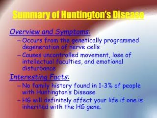
Overview of Huntington Disease (HD) • Hereditary degenerative neurological disease that leads to dementia. • Named after George Huntington, who first described it in 1872. DR. GEORGE HUNTINGTON(1850-1916)
Overview of Huntington Disease (HD) • Progressive dementia with involuntary movements, emotional disturbances, and metal deterioration. • Disease progresses slowly and death is usually due to an intercurrent infection. • Signs and symptoms develop in middle age, men and women are equally likely to develop the disease.
What Causes HD? • Genetically programmed degeneration of neurons. • Specifically affected are cells of the basal ganglia. • Structure that have many important functions, including coordinating movement. 1 Cerebrum2 Basal Ganglia3 Brain stem4 Cerebellum
What Causes HD? • All the characteristics can ultimately be traced back to a small change in the huntingtin gene on chromosome 4. • People who have Huntington disease have a larger huntingtin gene. This is due to a repetition in the code C-A-G. • Each C-A-G sequence codes for the amino acid glutamine, a protein building block. • People with HD simply have an increased number of these C-A-G repeats toward the beginning of their Huntington gene, coding for an excess number of glutamines in their huntingtin protein.
How is HD Inherited? • Genes are composed of deoxyribonucleic acid (DNA), a molecule shaped like a spiral ladder. • Each rung of the ladder is composed of base pairs. • There are four types of bases: adenine (A), thymine (T), cytosine (C), and guanine (G).
How is HD Inherited? • The gene that produces HD lies on chromosome 4. • Autosomal dominant disorder. • Only one copy of the defective gene is necessary to produce the disease. • The genetic defect responsible of HD is a repetition in the base pair (CAG) sequences of chromosome 4.
How is HD Inherited? • Each child of an HD parent has a 50-50 chance of inheriting the HD gene. • If a child does not inherit the HD gene, he or she will not develop the disease and cannot pass it to subsequent generations. • A person who inherits the HD gene will develop the disease in the middle age. • A small number of cases are sporadic, occurring even though there is no familiar history of the disorder. • These cases are caused by a genetic mutation that occurs during sperm development and that brings the number of CAG repeats into the range that causes the disease.
What Is the Function of the Huntingtin protein? • It plays a critical role in nerve cell function. Huntingtin regularly interacts with proteins found only in the brain. • People without Huntington have between 9 and 35 CAG repeats. • People with Huntington have between 36 and 121 CAG repeats. • Excess CAG repeat in the gene changes the shape of the protein (huntingtin). • Each CAG repeat codes for a specific amino acid – glutamine. • The extended number of glutamine repeats characterizes HD as one of nine polyglutamine expansion disorders**. • **Diseases which arise from extra copies of the CAG codon in certain segments of the DNA. The CAG codon codes for the amino acid glutamine.
How can the structure of huntingtin be altered? • If there are 41 CAG repeats in a row inside the gene, there will be 41 glutamines attached to each other in a row inside the huntingtin protein. • The production of multiple glutamines causes mutant huntingtin to fold into a different shape than normal. • Mutant huntingtin interacts with all sorts of other proteins that might normally be ignored, particularly in the spiny neurons deep inside the brain.
Huntingtin and Neurons • Excess huntingtin affects nerve cells’ dendrites. It causes dendrites to grow out of control. • Dendrites are found on every nerve cell and extend out from the cell body and are responsible for receiving and sending messages to other nerve cells. • New incomplete branches form and other branches become contorted, also causes dendrites to swell, break off, or disappear altogether. • This all contributes to nerve cell death. One method of cell death results from the release of excess glutamate
Counting CAG repeats in the Huntington’s disease gene by restriction endonuclease EcoP15I cleavage. Möncke-Buchner, E., S. Reich, M. Mücke, M. Reuter, W. Messer, E.E. Wanker, and D. H. Krüger. 2002. Counting CAG repeats in the Huntington’s disease gene by restriction endonuclease EcoP15I cleavage. Nucleic Acid Research 30: e83
Study Objective • Möncke-Buchner, et al. were interested in studying the numbers of CAG repeats in the HD gene by electrophoresis, and the cleavage after the insertion of EcoP15I. • Use of more advanced methods compared to the PCR fragments were deletions and insertions of the gene occur during the test. • For example Williams et al. found that amplifications products of the IT15 gene were greater with the use of capillary electrophoresis that using slab gel electrophoresis.
Fig. 1A - Recognition and cleavage sites of EcoP15I • Recognition sequence for EcoP15I consisting of 2 copies of the asymmetric sequence 5’-CAGCAG present in opposite strands of the DNA double helix. Showing that cleavage occurs 25-27 bp downstream of one of these sites. • This test was done in order to analyze the EcoP15I cleavage of HD in exon1. • Offers new advantages over existing procedures for the quantification of CAG repeats. Exhibits high-resolution quality, is not hazardous, prevents isotopic waste, and is time saving.
What is EcoP15I? • Type III restriction-modification enzyme. • Hetero-oligomeric proteins that behave as molecular machinesin response to their target sequences. • They translocate DNAin a process dependent on the hydrolysis of a nucleoside triphosphate. • For the ATP-dependent type III restriction andmodification systems, the collision of translocating complexestriggers hydrolysis of phosphodiester bonds in unmodified DNAto generate double-strand breaks. • Type I endonucleases breakthe DNA at unspecified sequences remote from the target sequence. • Type III endonucleases at a fixed position close to the targetsequence. • Notable for effective post-translational controlof their endonuclease activity.
Fig. 1B - Used DNA substrate at a distance of 72 bp. • Cleavage reactions were analyzed by polyacrylamide gel electrophoresis. • EcoP15I cleavage requires the presence of 2 5’-CAGCAG sequences that are located on opposite strands of the DNA. • A single inverse EcoP15I recognition site is located on the opposite strand, termed ‘i’ at 72 bp downstream of the CAG repeat. • Arrows indicate the EcoP15I cleavage sites 25-27 bp downstream of the various recognition sites.
Fig. 1A and Fig. 1B • In fig. 1A EcoP15I cleaves 25-27 bp downstream of one of the 2 recognition sites. • In fig. 1B EcoP15I recognition site is located on the opposite strand, termed ‘i’ at 72 bp downstream of the CAG repeat. • In the case of exon1 cleavage can either occur in the inverse site ‘i’ or 25-27 bp downstream of any of the (n-1) recognition sequences formed by the n CAG repeats.
Fig. 1C - Calculated length of the DNA fragments. • Calculated lengths of EcoP15I cleavage products expected for substrates fCAG30, fCAG35, and fCAG81.
Fig. 1D – Cleavage patterns • 10-fold excess of EcoP15I were added. • Cleavage generated characteristic ladders of DNA fragment. • Besides finding out that EcoP15I cleavages at the ‘i’ site, they also found that multiple DNA fragments generated by cleaving within the CAG repeat. • This means that EcoP15I recognizes and cleaves the multiple overlapping sites within the CAG repeat of HD in exon1 generating ladders of DNA fragments of different lengths.
Fig. 2 – Influence of EcoP15I concentration on cleavage efficiency and pattern. • This test was perform in order to determine whether the concentration of EcoP15I can influence the cleavage efficiency. • They found that by increasing the enzyme to substrate ratio enhanced the efficiency of cleavage. • But as we could see in lane 4 the presence does not correspond with the findings. • CAG repeats proximal to the inverse site were preferentially cleaved. Lane 1 – without EcoP15I Lane 2 – EcoP15I molar ration of 1 fmol Lane 3 – EcoP15I molar ratio of 10 fmol Lane 4 – EcoP15I molar ration of 20 fmol
ALFexpress DNA Analysis System • Since the ratio of EcoP15I enzyme to DNA substrate was too small to analyze, they used a high-resolution method called ALFexpress DNA Analysis System to separate and detect EcoP15I cleavage fragments. • ALFexpress DNA Analysis System is used for high performance and reliability in DNA analysis. • Employs a single-dye chemistry format in which Cy5 is used to label DNA fragments for detection. • Supported by a wide range of software, sequencing and genotyping reagents, and convenient application kits.
Fig. 3 - EcoP15I cleavage using ALFexpress DNA Analysis System • As mentioned before EcoP15I cleaved at the inverse ‘i’ recognition site of EcoP15I and at the recognition site or 25-27 bp downstream of any of the (n-1) recognition sequences formed by the n CAG repeats. • With the use of the ALFexpress DNA Analysis System they were able to count 29 DNA fragments in the ladder. • This number corresponds to the 29 overlapping EcoP15I recognition sites within the 30 CAG repeats of fCAG30.
Fig. 3 (cont.) • This demonstrates that all EcoP15I recognition sites are recognized and cleaved by EcoP15I. • They were able to determine the exact CAG repeat number.
Fig. 4 – Counting of CAG repeats using ALFexpress DNA Analysis System • Test performed in order to determine the repeat lengths in the borderline (30-39) and pathological range (more than 39). • They used 3 substrates (fCAG30, fCAG35 and fCAG81) to study the cleavage pattern. • Cleavage by EcoP15I resulted in a ladder of DNA fragments.
Fig. 4 (cont.) • EcoP15I cleaved at all 3 DNA substrates at the inverse EcoP15I recognition site and at each of the overlapping EcoP15I recognition sites 5’-CAGCAG within the CAG repeats. • Concluding this test we can say that EcoP15I preferred recognition sites located proximal to the inverse recognition site over more distant ones. • This preference is more likely caused by the faster formation of the active enzyme DNA complex when 2 EcoP15I recognition sites are located close to each other. • Exact quantification of the CAG repeat numbers in the normal as well as in the pathological range are necessary in order to determine the number of glutamine repeats characteristic of HD as one of nine polyglutamine expansion disorders.
Application • The count of CAG repeats is applicable to the analysis of further hereditary neurodegenerative diseases caused by expanded CAG repeats.
Prevalence • In the United States about 30,000 people have HD. • Prevalence is 1 in every 10,000 persons. • At least 150,000 others have a 50% risk of developing the disease. • HD appears to be less common in Japan, China, Finland, and African blacks.
Management • There is no cure to stop or reverse the curse of the disease. • Medication may be used to treat psychosis, depression, or movement problems. • Antipsychotic drugs may help to alleviate choreic movements and may also be used to help control hallucinations, delusions, and violent outbursts. • Tranquilizers can help control anxiety, depressions, pathological excitement and sever mood swings.