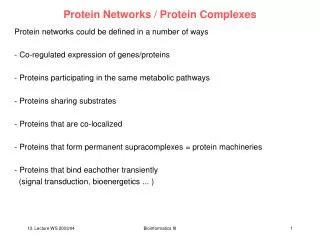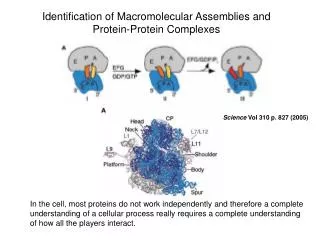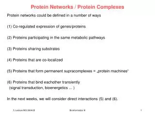
Intracellular calcium release channels as multi-protein complexes
350 likes | 521 Vues
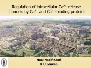
Intracellular calcium release channels as multi-protein complexes. Jan B. Parys K.U.Leuven. 7 th ECS meeting, June 13th, 2002. Intracellular Ca 2+ release channels: IP 3 Rs and RyRs. (Furuichi & Mikoshiba, 1995). Plasma membrane proteins. Cytoskeletal proteins. Homer. DHPR. Trp’s.

Intracellular calcium release channels as multi-protein complexes
E N D
Presentation Transcript
Intracellular calcium release channels as multi-protein complexes Jan B. Parys K.U.Leuven 7th ECS meeting, June 13th, 2002
Intracellular Ca2+ release channels: IP3Rs and RyRs (Furuichi & Mikoshiba, 1995)
Plasma membrane proteins Cytoskeletal proteins Homer DHPR Trp’s Ankyrin Vinculin Talin Cytosolic proteins G-proteins Alpha-actin Myosin II CaM CaBPs Sorcin S100 Kinase anchor proteins IRAG Calsequestrin Triadin Calreticulin FKBP’s Junctin Annexin VI ERmembrane proteins Chromogranins A and B Intraluminal proteins IP3R1IP3R2IP3R3 RyR1RyR2RyR3
What is the relation between the IP3R and the FKBP-type immunophilins ? • What is the relation between the IP3R and calmodulin ? • What is the relation between the IP3R and the cytoskeletal elements ?
FKBP12 and FKBP12.6 • 12-kDa FK506-binding protein • Peptidylprolyl isomerase activity ( FK506) • Immunophilins (+FK506) inhibit calcineurin Huse et al., 1999 • Modulators of the ryanodine receptors ( FK506) Stabilization of the channel Promotion of coupled gating • Modulators of the IP3 receptors ?
FKBP12-binding site Y2H IP3R1 VCTEGKNVYTEIKCNSLLPLDDIVRVVTHEDCIPEV IP3R2 ACTEGKNVYTEIKCNSLLPLDDIVRVVTHDDCIPEV IP3R3 ACAEGKNVYTEIKCTSLVPLEDVVSVVTHEDCITEV RyR1 QAGKGEALRIRAILRSLVPlDDLVGIISLPLQIPTG RyR2 HAGKGEAIRIRSILRSLIPLGDLVGVISIAFQMPTI RyR3 QTGKGEAIRIRSILRSLVPTEDLVGIISIPLKLPSL
SI FKBP12-binding site Ca2+ NH2 IP3 Ca2+ SIII Ca2+ Ca2+ FKBP12 Ca2+ Ca2+ P P ATP COOH ATP Ca2+ SII PORE Cytosol Lumen of the store IP3R1 VCTEGKNVYTEIKCNSLLPLDDIVRVVTHEDCIPEV IP3R2 ACTEGKNVYTEIKCNSLLPLDDIVRVVTHDDCIPEV IP3R3 ACAEGKNVYTEIKCTSLVPLEDVVSVVTHEDCITEV RyR1 QAGKGEALRIRAILRSLVPlDDLVGIISLPLQIPTG RyR2 HAGKGEAIRIRSILRSLIPLGDLVGVISIAFQMPTI RyR3 QTGKGEAIRIRSILRSLVPTEDLVGIISIPLKLPSL Ca2+
SH-SY5Y BC3H1 FK506 and FKBP12 affect Ca2+ releasethrough the RyRs but not through the IP3Rs
A7r5: IP3R1 IP3R3 Sf9: IP3R1 IP3R3 Cerebellum: IP3R1 RyR1 Retention of the intracellular Ca2+-release channels by GST-FKBP12 affinity assay 2 1 s e e P t a B m z K o i l s i F b - o T T r u l c S S i o M G G S RyR1 (Skel. muscle) RyR3 (HEK293)
Mutational analysis of FKBP12-binding site RyR3 IRIRSILRSLVPTEDLVGIISIP RyR3/V2322L IRIRSILRSLLPTEDLVGIISIP (as in IP3R1) RyR3/V2322I IRIRSILRSLIPTEDLVGIISIP (as in RyR2) RyR3/V2322D IRIRSILRSLDPTEDLVGIISIP (negat. charge) RyR3/IP3R1 IRIRSICNSLLPLDDIVGIISIP (as in IP3R1)
Putative model RyRs : XP residue as an -helix breaker IP3Rs : XP residue in a different secondary structure FKBP12 : binding protein binding stable interaction FKBP12 : enzyme catalysis transient interaction
RyRs and IP3Rs have differentFKBP-binding properties • FKBP12-binding site on the IP3R is much less efficient than the homologous site in the RyR. • Higher-order structure may be important. • RyRs: stable interaction FKBP12 is regulatory subunit. • IP3Rs: weak or transient interaction chaperone function? effects on kinetics? • FK506 can act on SERCA and on calcineurin.
What is the relation between the IP3R and the FKBP-type immunophilins ? • What is the relation between the IP3R and calmodulin ? • What is the relation between the IP3R and the cytoskeletal elements ?
ApoCaM: Ca2+/CaM: ACTIVATOR OF RYR1 INHIBITOR OF RYR1 Effects on RyR 3614 3643 Ca2+ (Rodney et al., 2001)
IP3R Effects of CaM on Ca2+ release: A7r5 (Missiaen et al., 1999)
IP3R Effects of CaM on Ca2+ release: 200 μM Ca2++10 μM CaM +20 μM CaM A7r5 (Missiaen et al., 1999) Cerebellum (Michikawa et al., 1999)
IP3R Effects of CaM on Ca2+ release: Effects of CaM on IP3 binding: 200 μM Ca2++10 μM CaM +20 μM CaM A7r5 (Missiaen et al., 1999) Cerebellum (Michikawa et al., 1999) Sf9 (Cardy & Taylor, 1998)
IP3R Effects of CaM on Ca2+ release: Effects of CaM on IP3 binding: 200 μM Ca2++10 μM CaM +20 μM CaM A7r5 (Missiaen et al., 1999) Cerebellum (Michikawa et al., 1999) Lbs-1 Lbs-2 Lbs-3 CaM (μM) Sf9 (Cardy & Taylor, 1998) Lbs-domains (Vanlingen et al., 2000)
SI CaM Ca2+ NH2 IP3 Ca2+ SIII Ca2+ Ca2+ Ca2+ Ca2+ CaM P P ATP CaM COOH ATP Ca2+ SII PORE Cytosol Lumen of the store Low-affinityCa2+-dependentNot in neuronalIP3R1 (SII+) Ca2+ CaM-binding sites High-affinityCa2+-dependentTypes 1 and 2
1 581 Lbs-1 226 581 Lbs-1 1-225 Effects of CaMon IP3 binding +HIS +HIS 100 80 60 [3H]IP3 binding (%) [3H]IP3 binding (%) 40 20 0 0.1 1 10 CaM1234 (μM) Ca2+ Control ApoCaM Ca2+ CaM CaM1234 Ca2+ CaM1234
1 581 Lbs-1 226 581 Lbs-1 1-225 Effects of CaMon IP3 binding +HIS +HIS 100 80 60 [3H]IP3 binding (%) [3H]IP3 binding (%) IC50~ 2 μM 40 20 0 0.1 1 10 CaM1234 (μM) Ca2+ Control ApoCaM Ca2+ CaM CaM1234 Ca2+ CaM1234
1 581 Lbs-1 226 581 Lbs-1 1-225 Cyt1 Cyt2 Detailed localisation of the N-terminal CaM-binding site +HIS +HIS +GST 1 159 154 309 Ca2+ CaM1234 EGTA
1 581 Lbs-1 226 581 Lbs-1 1-225 Cyt1 Cyt2 Detailed localisation of the N-terminal CaM-binding site +HIS +HIS +GST 1 159 154 309 1-5-10 1-5-10 53% IQ 1-5-8-14 76% IQ 70% IQ A C D B E F 1.0 2+ 200 µM free Ca 0.8 • Band-shift experiments on non-denaturing gels • Interaction with dansyl-CaM 1 mM EGTA 0.6 0.4 0.2 0.0 -0.2 A B C D E F
1 581 Lbs-1 226 581 Lbs-1 1-225 Cyt1 Cyt2 Detailed localisation of the N-terminal CaM-binding site +HIS +HIS +GST 1 159 154 309 1-5-10 1-5-10 53% IQ 1-5-8-14 76% IQ 70% IQ A C D B E F 1.0 200 µM Ca2+1mM EGTA Kd 0.1 μM Kd 1 μM 0.8 0.6 0.4 Intensity loss (1-B/Bo) 0.2 0.0 -0.2 A B C D E F
CaM1234 does not inhibit IP3-induced Ca2+ release IP3 1 μM IP3 1 μM + CaM 10 μM IP3 1 μM + CaM1234 10 μM
N-terminal CaM-binding site of the IP3R • Discontinuous (aa 49-81 and 106-128). • Low affinity. • Ca2+ independent. • Involved in inhibition of IP3 binding. • Not involved in inhibition of IP3-induced Ca2+ release. • Other possible functions: Role in conformation of the IP3R ? Tethering of CaM ? Binding site for CaM-like proteins ? CaBP-1? Protective effect ? Proteolysis? Oxidative stress?
What is the relation between the IP3R and the FKBP-type immunophilins ? • What is the relation between the IP3R and calmodulin ? • What is the relation between the IP3R and the cytoskeletal elements ?
Localization of IP3R1 and IP3R3in A7r5 smooth-muscle cells IP3R1 IP3R3
AVP > 1h ImipramineIP3-ester ThapsigarginCPA [Ca2+]cyt Redistribution of IP3R1 after prolonged stimulation Resting cells + AVP
Percentages of cells with: perinuclear IP3R1 cytoplasmic IP3R1 AVP AVP OAG OAG Control Control TG+Staur. TG+Staur. AVP+Staur. AVP+Staur. AVP+BIM AVP+BIM Role of PKC 100 80 60 Cells (%) 40 20 0
Role of cytoskeleton Percentages of cells with: Resting cells perinuclear IP3R1cytoplasmic IP3R1 +AVP AVP AVP Control Control AVP+Nocod. AVP+Nocod. Microtubular network 80 70 60 50 Cells (%) 40 30 20 10 0
Various IP3R isoforms can have a different distribution. The localization of the IP3Rs is dynamic and change with the physiological status of the cell. PKC activation seems an important determinant of the intracellular localization pattern. The microtubular network is involved but which protein(s) determine the IP3R localization is not yet ascertained. Determinants of the localization of IP3Rs
Conclusions • The intracellular Ca2+ release channels IP3Rs and RyRs act as part of a multiprotein complex. • Many proteins have been proposed as partners. • The existence of consensus binding sites is not sufficient for assuring binding in vivo. Higher-order structure, experimental conditions and indirect associations can be involved. • RyRs and IP3Rs are regulated by different proteins. • Future work on the IP3R will focus on the interactions with the various Ca2+-binding proteins and with the different scaffolding proteins.
IP3-team (Leuven, Belgium) Geert Bultynck Patrick De Smet Nael Nadif Kasri Ilse Sienaert Karolina Szlufcik Kristel Van Acker Sara Vanlingen Elke Vermassen Geert Callewaert Humbert De Smedt Ludwig Missiaen Jan B. Parys
IP3-team (Leuven, Belgium) Geert Bultynck Patrick De Smet Nael Nadif Kasri Ilse Sienaert Karolina Szlufcik Kristel Van Acker Sara Vanlingen Elke Vermassen Geert Callewaert Humbert De Smedt Ludwig Missiaen Jan B. Parys