Download

1 / 45
520 likes | 1.51k Vues
Non-invasive Positive Pressure Ventilation. Dr.jarahzadeh Intensivist. Introdaction * Nippv is recent phenomenon, mainly because of advances in noninvasive interfaces and ventilator modes * NIPPV delivered o2 by nasal or oronasal mask * The efficacy of noninvasive positive-pressure

E N D
Non-invasive Positive Pressure Ventilation Dr.jarahzadeh Intensivist
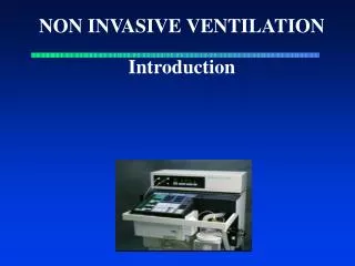
Introdaction * Nippv is recent phenomenon, mainly because of advances in noninvasive interfaces and ventilator modes * NIPPV delivered o2 by nasal or oronasal mask * The efficacy of noninvasive positive-pressure Ventilation has been demonstrated for acute pulmonary edema, for respiratory failure in immunocompromised patients, and to facilitate extubation in COPD patients.
* Patients who develop respiratory failure or who refuse intubation are potentially good candidates for noninvasive positive-pressure ventilation *Several factors are vital to the success of noninvasive positive-pressure ventilation: careful patient selection; properly timed initiation; comfortable, well-fitting interface; coaching and encouragement; and careful monitoring. *Noninvasive ventilation should be used to avert endotracheal intubation rather than as an alternative to it.
Definition The application of positive pressure ventilation without using an endotracheal tube. or As the provision of ventilatory assistance to the lungs without an invasive artificial airway
History With the introduction of nasal CPAP to treat obstructive sleep apnea in the early 1980s, NIPPV rapidly displaced negative-pressure ventilation as the treatment of choice for chronic respiratory failure in patients with neuromuscular and chest wall deformitie The past 12 years, noninvasive ventilation has moved from the outpatient to the inpatient setting, where it is used to treat acute respiratory failure. Until the early 1960s, negative-pressure ventilation in the form of tank ventilators was the most common type of mechanical ventilation outside the anesthesia suite
Non-invasiveVentialtion 1- Positive pressure 2-Negative Pressure
Advantage(NIPPV) * lowers morbidity and mortality * Shorten hospital length of stay, thus reducing costs. *Decreased direct upper airway trauma & bypass of the upper airway defense mechanisms *Allows patients to eat orally, vocalize normally, and expectorate secretions. * Noninvasive ventilation reduces infectious hospital including pneumonia,sinusitis, and sepsis.
Goals of NIV • Relieve symptoms • Reduce work of breathing • Offset the effect of iPEEP • Improve gas exchange • Minimize risk of barotrauma • Avoid intubation
Indication Airway Obstruction COPD Asthma Cystic fibrosis Obstructive sleep apnea or obesity hypoventilation Upper airway obstruction Facilitation of weaning in COPD Extubation failure in COPD
Indication HypoxemicRespiratory Failure ARDS Pneumonia Trauma or burns Acute pulmonary edema (use of CPAP) Immuno compromised patients Restrictive thoracic disorders Post operative patients Do-not-intubate patients During bronchoscopy
Respiratory arrest Medically unstable Unconscious, unable to protect airways Excessive secretions Significant vomiting Agitated or uncooperative Facial trauma, burns, surgery or anatomic abnormalities interfering with mask application Exclusion Criteria
PATIENT SELECTION Primary-step Identify patients in need of ventilatory assistance by using clinical and blood gas criteria. Good candidates are those with moderate to severe dyspnea, tachypnea, and impending respiratory muscle fatigue( use of accessory muscles of breathing or abdominal paradox). The level of tachypnea ( COPD when the respiratory rate exceeds 24 breaths per minute & hypoxemic respiratory failure, higher respiratory rates are used, in the range of 30 to 35 breaths per minute.
second step Exclude patients for whom noninvasive ventilation would be unsafe. Those with frank or imminent respiratory arrest Patients who are medically unstable with hypotensive shock, uncontrolled upper gastrointestinal bleeding, unstable arrhythmias, or life-threatening ischemia . who are uncooperative, unable to adequately protect their upper airway or clear
PREDICTORS OF SUCCESS DURING ACUTE APPLICATIONS OF NPPV • Younger age • Lower acuity of illness (APACHE score) • Able to cooperate; better neurologic score • Able to coordinate breathing with ventilator • Less air leaking, intact dentition • Hypercarbia, but not too severe (PaCO2 > 45 mm Hg, < 92 mm Hg) • Acidemia, but not too severe (pH < 7.35, > 7.10) • Improvements in gas exchange and pulse and respiratory rates within first 1-2 h
INITIATION OF NONINVASIVE VENTILATION 1-Appropriate candidate selected, 2-Ventilator and interface must be chosen, 3-Ventilator settings must be selected, 4-Location ( Icu or step-down unit that offers adequate continuous monitoring until stabilized)
Comporison of Noninvasive mechanical ventilators with standard critical care ventilators NIMV offers a more portable technology due to the reduced size of the air compressor. Because of this reduction in size, these noninvasive ventilators do not develop pressures as high as their critical care ventilator counterparts. (>30 cm H20) Noninvasive ventilators have a single-limb tubing circuit that delivers oxygen to the patient and allows for exhalation. lack oxygen blenders or sophisticated alarm or battery backup systems
Modes of Noninvasive Mechanical Ventilation 1-Pressure modes 2-volume modes * volume ventilation, initial tidal volumes range from 10 to 15 mL.kg. Pressure-cycled vents are better tolerated than volume-cycled vents
Pressure modes **Continuous Positive Airway Pressure(CPAP) Continuous positive airway pressure (CPAP) is a mode for invasive and noninvasive mechanical ventilation. It provides positive airway pressure throughout the respiratory cycle. This static, positive pressure is maintained constantly during inhalation and exhalation CPAP is not a stand-alone mode of assisted mechanical ventilation. It is equivalent to positive end-expiratory pressure (PEEP) and facilitates inhalation by reducing pressure thresholds to initiate airflow. This mode should never be used in patients who may have apneic episodes because of the lack of a backup rate.
Pressure modes Spontaneous Modes In spontaneous mode, the airway pressure cycles between an inspiratory positive airway pressure (IPAP) and an expiratory positive airway pressure (EPAP). This is commonly referred to as bilevel or biphasic positive airway pressure (BL-PAP or BiPAP). The patient's inspiratory effort triggers the switch from EPAP to IPAP. The limit during inspiration is the set level of IPAP. The inspiratory phase cycles off, and the machine switches back to EPAP when it detects a cessation of patient effort, indicated by a decrease in inspiratory flow rate, or a maximum inspiratory time is reached, typically 2-3 seconds. Tidal volume (Vt) varies breath to breath and is determined by degree of IPAP, patient effort, and lung compliance. Spontaneous mode depends on patient effort to trigger inhalation. A patient breathing at a low rate can develop a respiratory acidosis.
Spontaneous/timed (ST) mode The trigger in the ST mode can be the patient's effort or an elapsed time interval, predetermined by a set respiratory backup rate. If the patient does not initiate a breath in the prescribed interval, then IPAP is triggered. For machine-generated breaths, the ventilator cycles back to EPAP based on a set inspiratory time. For patient-initiated breaths, the ventilator cycles as it would in the spontaneous mode. Pressure modes
Conceptually: One can consider BiPAP as PEEP with pressure support (PS). The pressure during the inspiratory phase is termed IPAP and is analogous to PS. The pressure during the expiratory phase is termed EPAP and is analogous to PEEP. The IPAP is necessarily set higher than EPAP by a minimum of 5cmH2O, and the difference between the two settings is equivalent to the amount of PS provided
Initiating Noninvasive Mechanical Ventilation Either a face mask or a nasal mask can be used, but a nasal mask is generally better tolerated. A respiratory therapist must measure the patient to ensure a good fit and seal. Initially supply 3 to 5 cm H2O of CPAP with supplemental oxygen. sequentially increase the CPAP pressure by 2 to 3 cm H2O increments every 5 to 10 minutes (ABG-Pulse oximetry) Recommended initial settings for BiPAP machines in the noninvasive support of patients in respiratory distress or failure are IPAP of 8 cm H2O and EPAP of 3 cm H2O, for a pressure support (IPAP minus EPAP) of 5 cm H2O. The level of supplemental oxygen flowing into the circuit should be governed by goal pulse oximetry and corroborated by ABG results as necessary; it is appropriate to initiate therapy with 2 to 5 L/minute, but this amount should be adjusted with each titration of IPAP or EPAP.
Conceptually: The intrinsic positive end-expiratory pressure (PEEPi), or auto-PEEP, cannot be measured by a noninvasive ventilator; therefore, EPAP should generally be maintained below 8 to 10 cm H2O to be certain that it does not exceed PEEPi in patients with obstructive lung disease. The IPAP must always be set higher than EPAP
Management Strategies • COPD • Main goal to decrease work of breathing (decreasing V/Q mismatch) and provide adequate ventilation • Relatively low EPAP: 5-8cm H2O (assuming no obesity or sleep disordered breathing) • Relatively moderate IPAP+EPAP: 10-14cm H2O • Goal to have at least a 5cm H2O differential between EPAP and IPAP+EPAP; may need to go higher depending on ventilation requirements • ieBiPAP 14/10 or 8/5
From a Cochrane Review • A meta-analysis of 14 studies of NIV in COPD exacerb showed: • ¯mortality ( RR 0.52 ) • ¯need for intubation ( RR 0.41 ) • ¯ pCO2, and resp rate faster • ¯length of stay by 3.24 days • ¯complications of treatments
Management Strategies • CHF • Goal is to decrease work of breathing, decrease afterload and decrease overall static pressure • Relatively moderate EPAP: 6-12 cm H2O • Relatively low IPAP+EPAP: 12-18cm H2O • Patient will benefit mostly with EPAP unless other concurrent disease ( COPD, Obesity-Hypoventilation) • Typical starting point: BiPAP 10/6
Management Strategies • Obesity-Hypoventilation Syndrome • Goal of therapy is to decrease work of breathing and increase ventilation • Combined disease as >90% will also have concurrent Obstraction sleep Apnea(OSA) • EPAP: usually on the higher side; enough to overcome OSA and cardiopulm disease: ~10cmH2O, more for bigger individuals • IPAP+EPAP: at least a 4cm H2O differential • Need to adjust according to ventilation requirements; may benefit from back up rate
Management Strategies • Sleep Disordered Breathing • Most often post-op with known OSA or as a complication associated with admit (CHF or Obesity-Hypoventilation) • For elective admit with known OSA: usual CPAP/BiPAP unless physiologic changes with acute illness, surgery or narcotics.
Management Strategies • Neuromuscular Disease • Goal to decrease work of breathing, decrease fatigue, assist ventilation • EPAP: usually low; 4-5cm H2O • IPAP+EPAP: at least 4cmH2O differential • May benefit from backup rate
Management Strategies • Other causes of respiratory failure • Pneumonia/ARDS • Cancer and respiratory failure • Post-op management • Settings depend on disease and other cardiopulmonary disease • Most often used as a bridge to mechanical ventilation or for pts DNR/DNI • Usually moderate settings: 12/8 or 14/8
Interface *Definition: The device that makes physical contact between the patient and the ventilator is termed the interface. Interfaces for NPPV come in a variety of shapes and sizes Include: Nasal mask,Nasal pillow, Oronasal mask (face mask) or the helmet. Ideally, interfaces should be comfortable, offer a good seal, minimize leak, and limit dead space.
Nasal masks are widely used for the administration of CPAP or noninvasive ventilation, particularly for chronic applications. Nasal masks are usually better tolerated than full face masks for long-term applications, because they cause less claustrophobia and discomfort and allow eating,conversation, and expectoration. The standard nasal mask is a triangular or cone-shaped clear plastic device that fits over the nose and uses a soft cuff that forms an air seal over the skin. Full facemasks cover both the nose and the mouth and are preferable to nasal masks in the acute setting.
The efficacy of both nasal and oronasal masks in lowering PaC02 and avoiding intubation is similar in the acute setting, but in a recent randomized, patients tolerated the full facemask better because of reduced air leakage through the mouth. Selection of a comfortable mask that fits properly is key to the success of noninvasive ventilation. The full facemask should be tried first in the acute setting, and if possible, The mask straps are then tightened with the least tension necessary to avoid excessive air leakage.
NIPPV masks • Nasal mask
NIPPV masks • Full face mask
NIPPV masks • Full face mask Most of our patients!!
NIPPV machines • BiPAP
NIPPV machines • CPAP machine
Head straps Head straps hold the mask in place and are important for patient comfort. Straps attach at two to five points, depending on the type of mask. More points of attachment add to stability.
OXYGENATION AND HUMIDIFICATION Oxygen is titrated to achieve a desired oxygen saturation, usually greater than 90% to 92% Either by using oxygen blenders on critical care and some bilevel ventilators or By adjusting liter flow (up to 15 L/min) delivered via oxygen tubing connected directly to the mask or ventilator circuit. Bilevel ventilators have limited oxygenation capabilities (maximal inspired oxygen fraction( %45 to 50) so ventilators with oxygen blenders should be used for patients with hypoxemic respiratory failure. A heated humidifier should be used to prevent drying of the nasal passage and oropharynx when the duration of application is anticipated to be more than a few hours.
MONITORING Once noninvasive ventilation is initiated, patients should be closely monitored in a critical care unit or a step-down unit until they are sufficiently stable to be moved to a regular medical floor. The aim of monitoring is Relief of symptoms, reduced work of breathing, improved or stable gas exchange, good patient-ventilator synchrony, and patient comfort A drop in the respiratory rate with improved oxygen saturation or improving pH with a lower PaCO2, reduce heart rate, within the first 1 to 2 hours portends a successful outcome. The absence of these propitious signs indicates a poor response to noninvasive ventilation
MONITORING OF PATIENTS RECEIVING NON-INVASIVE VENTILATION IN ACUTE CARE SETTINGS Location Critical care or step-down unit Medical or surgical ward if able to breathe unassisted for >20-30 min "Eyeball“ test Dyspnea Comfort (mask, air pressure) Anxiety Asynchrony Leaks Vital signs Respiratory and heart rates Blood pressure Continuous electrocardiography Gas exchange Continuous oximetry Arterial blood gases (baseline after 2 h ,and as clinically indicated)
ADVERSE EFFECTS AND COMPLICATIONS in NIV The mask, Discomfort and erythema or skin ulcers. Airflow or pressure, Conjunctival irritation. Ear pain. nasal or oral dryness . Nasal congestion and discharge. Gastric insufflation. Patient-ventilator asynchrony Caused by high airflow is usually indicative of air leaking through the mouth.