
SlideServe
Menu
Préférences
© SlideServe · v2026

Classification of Cellular Organism ( according to cell structure)

catriona avatar
19 diapositives · 980 Views · Nov 07, 2012

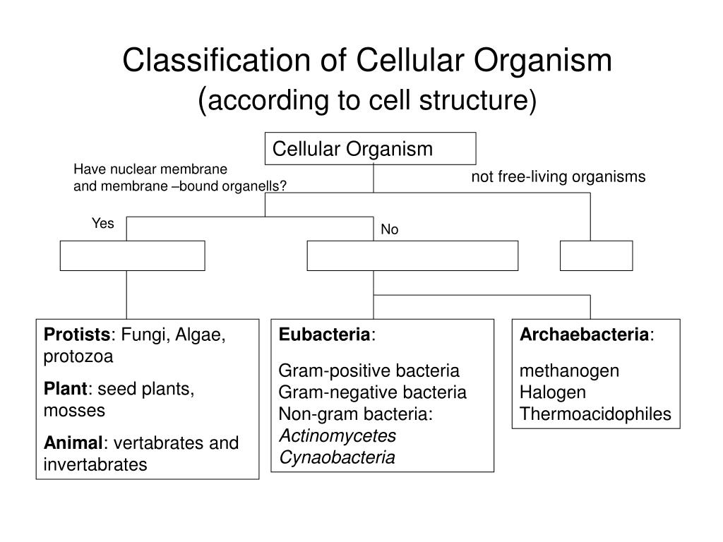
Classification of Cellular Organism ( according to cell structure). Cellular Organism. Have nuclear membrane and membrane –bound organells?. not free-living organisms . Yes. No. Protists : Fungi, Algae, protozoa Plant : seed plants, mosses Animal : vertabrates and invertabrates .
Classification of Cellular Organism ( according to cell structure) - slide 1
01 / 19Classification of Cellular Organism ( according to cell structure)
1 / 17

Télécharger la présentation

Classification of Cellular Organism ( according to cell structure)
Une image / un lien ci-dessous est fourni (tel quel) pour télécharger la présentation

Politique de téléchargement : le contenu du site vous est fourni TEL QUEL pour votre information et votre usage personnel ; il ne peut être vendu, concédé sous licence ni partagé sur d’autres sites sans l’accord de son auteur.Le contenu vous est fourni TEL QUEL, pour votre information et votre usage personnel uniquement. Téléchargez la présentation en cliquant sur ce lien. Lors du téléchargement, si vous ne parvenez pas à télécharger une présentation, l’éditeur a peut-être supprimé le fichier de son serveur.Pendant le téléchargement, si vous ne parvenez pas à obtenir une présentation, le fichier a peut-être été supprimé par l’éditeur.

F I N

Plus de contenu associé

Transcription de la présentation
  1. Classification of Cellular Organism(according to cell structure) Cellular Organism Have nuclear membrane and membrane –bound organells? not free-living organisms Yes No Protists: Fungi, Algae, protozoa Plant: seed plants, mosses Animal: vertabrates and invertabrates Eubacteria: Gram-positive bacteria Gram-negative bacteria Non-gram bacteria: Actinomycetes Cynaobacteria Archaebacteria: methanogen Halogen Thermoacidophiles

  2. Classification of Cellular Organism(according to cell structure) Cellular Organism Have nuclear membrane and membrane –bound organells? not free-living organisms Yes No Protists: Fungi, Algae, protozoa Plant: seed plants, ferns, mosses Animal: vertabrates and invertabrates Eubacteria: Gram-positive bacteria Gram-negative bacteria Non-gram bacteria Actinomycetes Cynaobacteria Archaebateria: methanogen Halogen Thermoacidophiles

  3. Procaryote Procaryotes have membrane around the cell genetic information and membrane-bound organelles • Bacteria: e.g. E. Coli, Rhodospirillum sp. • Size: • Grow rapidly: e.g. one cell can replicate into over a million cells in just 12 hours. In contrast, a human cell takes 24 hours to split. • Utilize carbon sources: carbohydrates, hydrocarbon, protein and CO2.

  4. Picture courtesy of http://micro.magnet.fsu.edu/cells/procaryotes/images/procaryote.jpg

  5. Procaryote Cell Structure Nuclear region There is around the nuclear region containing genetic materials such as chromosomes and DNA (deoxyribonucleic acid). Chromosomes: A chromosome is, , which contains many genes, regulatory elements and other intervening nucleotide sequences. The DNA which carries genetic information in biological cells is normally packaged in the chromosomes.

  6. http://micro.magnet.fsu.edu/cells/procaryotes/images/procaryote.jpghttp://micro.magnet.fsu.edu/cells/procaryotes/images/procaryote.jpg

  7. Procaryote Cell Structure Cytoplasm In cytoplasm, there are some visible structures: • : sites of protein synthesis, 10,000 per cell, 10 -20 nm, 63% RNA and 37% protein. • : source of key metabolites, containing polysaccharides, lipids and sulfur granules. Sizes vary between 0.5-1 µm. • :DNA molecules separate from the chromosomal DNA and capable of autonomous replication. Usually occur in bacteria. e.g E.coli Application in Genetic Engineering.

  8. http://micro.magnet.fsu.edu/cells/procaryotes/images/procaryote.jpghttp://micro.magnet.fsu.edu/cells/procaryotes/images/procaryote.jpg

  9. Procaryote Cell Structure Cytoplasmic membrane • The cytoplasm is surrounded by a membrane called cytoplasmic membrane. • The cytoplasmic membrane contains 50% protein, 30% lipids and 20% carbohydrates.

  10. http://micro.magnet.fsu.edu/cells/procaryotes/images/procaryote.jpghttp://micro.magnet.fsu.edu/cells/procaryotes/images/procaryote.jpg

  11. Procaryote Cell Structure Cell wall - Eubacteria cell walls contain lipids & peptidoglycan which is a complex polysaccharide with amino acids and forms a structure somewhat like chain-link fence. - Archaebacteria cell walls do not have peptidoglycan. Outer membrane: Some bacteria (gram negative cells) have. - -

  12. http://micro.magnet.fsu.edu/cells/procaryotes/images/procaryote.jpghttp://micro.magnet.fsu.edu/cells/procaryotes/images/procaryote.jpg

  13. Procaryote Cell Structure Capsule: Extracellular products can adhere to or become incorporated within the surface of the cell. Certain cells have a coating outside the cell wall called capsule. It contains polysaccharides or polypeptide and forms biofilm response to environmental challenges. Flagellum: is for cell motion. Pilus (Pili, pl.) A pilus is a hairlike structure on the surface of a cell. Pili enable the transfer of plasmids between the bacteria. An exchanged plasmid can add new functions to a bacterium, e.g., an antibiotic resistance.

  14. Procaryotes Procaryotes include - -

  15. Eubacteria Cell chemistry of eubacteria is similar to eucaryotes. Classification Gram stain: Hans Christian Gram in 1884 developed the technique of gram stain which has been used to classify the eubacteria. Gram staining procedure: Fixing the cells by heating Dye with crystal violet – stain purple Iodine and ethanol are added a. : The cells are colorless after Gram staining procedure. Gram-negative organisms will be counterstained with safranin and appear red or pink. Such cells membrane supported by peptidoglycan e.g. E. coli. b. : The cells remain purple after gram staining and counterstaining procedures. Such cells outer membrane but with a rigid cell wall and thick peptidoglycan layer, e.g. B. subtilis. http://student.ccbcmd.edu/courses/bio141/labmanua/lab6/images/gram_stain_11.swf

  16. Eubacteria Other types of eubacteria: • Non gram bacteria: some bacteria are not gram-positive or negative. e.g Mycoplasma is non gram bacteria lack of cell wall. It is an important cause of peumonia and other respiratory disorders. Actinomycetes: bacteria but, morphologically resembles molds with their long and high branched hyphae. They are important source of antibiotics.

  17. Archaebacteria Archaebacteria cells differ greatly from eubacteria at the molecular level. - no peptidoglycan - The nucleotide sequences in the ribosomal RNA are similar within the archaebacteria but distinctly different from eubacteria. - The lipid composition of the cytoplasm membrane is very different for the two groups. This category includes: - methanogen: methane-producing bacteria - Halogen: living only in very strong salt solutions - Thermoacidophile: growing at high temperatures and low pH.

  18. Procaryote Reproduction Reproduction: exclusively asexual through The chromosome is duplicated and attaches to the cell membrane, and then the cell divides into two equal cells.

  19. Binary Fission http://www.beyondbooks.com/lif72/2a.asp