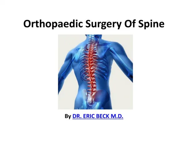

PROBLEMS DURING ORTHOPAEDIC SURGERY
E N D
Presentation Transcript
PROBLEMS DURING ORTHOPAEDIC SURGERY Dr. M.J. MAHANTHESHA SHARMA M.D., D.A., PROFESSOR DEPARTMENT OF ANAESTHESIA J.J.M .MEDICAL COLLEGE DAVANGERE – 577 004.
PROBLEMS DURING ORTHOPAEDIC SURGERY • Air way problems • Positioning related problems • Blood loss related problems • Bradycardia / Asystole • Paraplegia during scoliosis surgery • Neuropraxia • DVT problem • Thromboembolism problems • Fat embolism • Bone cement related problems • Anticoagulation therapy • Tourniquet problems • Postoperative delirium and confusion
AIRWAY PROBLEMS • Complex airway challenges are common • Juvenile rheumatoid arthritis, ankylosing spondylitis, prior cervical fusion. • Impossible to intubate with conventional laryngoscopy. • Failed intubation, trauma to airway, respiratory distress after extubation.
PROBLEMS • Rheumatoid arthritis C1-2 subluxation – instability Uncontrolled flexion – compromise the spinal cord. • Uncontrolled flexion during spinal surgery-quadriplegia. • Athletic patients coming for sport related surgery - Acute respiratory distress after extubation. - Low pressure pulmonary edema. • Cricoarytenoid joints – decreases the glottic area. • Intrinsic and extrinsic airway diseases - PFT.
PREVENTION &MANAGEMENT • Careful assessment of the airway • Selection of regional technique • Select fibroptic technique under light sedation. • Careful positioning them for surgery. • If GA is required use fibroptic bronchoscope. • Check neurological functions • Acute respiratory distress after extubation prevented by – • Fibroptic intubation • Kept intubated 4-5 hours, head elevation 30°. • Use smaller endotracheal tubes.
POSITION RELATED PROBLEMS • Requires different intraoperative positions. • Limbs are placed in unphysiological positions. • Pressure sores – pressure effect. • Nerve injury - compression or stretch. • Ischaemia – vascular kink or obstruction. • Ischaemia or compartment syndrome results. • Avoid active movement of Ankylosed joint.
SPECIFIC PROBLEMS DUE TO POSITIONING • THR (dependent limb) – compartment syndrome. • Spinal surgery - prone - Brachial plexus palsy • Prone - compression - femoral or lateral cutaneous nerves. • Prone – compression of eye – Post op. blindness. • Brachial plexus stretch – Palsy - shoulder arthroplasty.
PREVENTION • Correct positioning, proper padding. • Avoid compression on eye. • Avoid unnecessary stretching of the limbs. • Avoid tight bandages and cast. • Care of abduction braces after shoulder surgery.
BLOOD LOSS PROBLEMS • Major Procedures likely to have estimated blood loss >1lt to 50% of blood volume • Revision total hip arthroplasty • Arthroplasty for congenital hip deformity • Removal of infected prosthesis • Revision IM nailing of a femur fracture • Resection and reconstruction of bone lesions • Bilateral total knee arthroplastis • Biopsy of any sacral lesion • Spinal fusion at more than three levels.
Hypotension • Main complication of blood loss • Induced hypotensive technique • Monitor intra op. SV and filling pressure. • Homologous transfusion • Intraoperative cell saver. • Preoperative autologus blood donation. • Invasive monitor – arterial pressure, CVP. • Preoperative haematocrit value.
Treatment of hypotension • Maintain haematocrit level • Volume replaced by • Crystalloids • Colloids • Blood and blood products. • Vesopressor • Administration of fluids by CVP. • Don’t overload in high risk patients.
Bradycardia/ Asystole • GA with vacuoronium / fentanyl combination. • Regional – severe acute bradycardia. • Common life threatening during regional. • Block above T4 decrease heart rate. • Needs beta agonists or atropine. • Bezold-Jarisch type of reflex even below T6 block. • Vagal mediated leads to asystole. • Triggered by reduction in intrathoracic volume. • Shoulder surgery - sitting - venous pooling volume.
Management • Rapid treatment is required. • Some times death or permanent brain damage. • Proper vigilance • Maintain adequate – blood volume with IV fluids • Prophylactic administration of atropine, beta agonists. • Treat with epidrine 10-20mg, atropine 0.4 – 0.8 mg. • Asystole treated by epinephrine, chest compression,
Paraplegia and scoliosis surgery • Tragedy, uncommon in uncomplicated cases. • Congenital scoliosis and more severe thoracic curves. • Spinal cord function monitor - SSEP and wakeup test. • Hypotensive anaesthesia with MAP 60 mm of Hg. • Facilitate optimal blood flow to spinal cord. • Stable blood volume with CVP and urine output. • Avoid massive blood loss. • Care during spinal distraction. • Maintain stable circulation. • Invasive monitoring as and required. • Blood transfusion as and required.
Neuropraxia • Postoperative nerve injuries are common. • Neuropathy, surgical injury, malpositioning or tourniquet. Prevention : • Avoid malpositioning, tight bandages or casts. • Avoid compartment syndrome. • Perioperative neuropraxia - anaesthesiologists concern. • Legally shared the responsibility with surgeon. • Medico legal problems are common. • Preoperative nerve function assessment documented.
DVT PROBLEMS • Complications of lower extremity surgeries. • Fatal PE is 1-2% without thrombosis prophylaxis. • Major trauma 58% of DVT, 15% proximal veins. • With prophylaxis – DVT reduced to 20%. • Fatal PE almost minimal or eliminated. • 15% of all postoperative deaths due to PE.
Thromboembolism • Hip and knee surgeries • Advanced age and Female sex • Previous history of thromboembolic disease • Malignant diseases • Prolonged bed rest / immobilization • General anaesthesia increased incidence. • Surgical or accidental trauma. • Fracture of femur and tibia high risk.
Pulmonary embolism • PE is not a disease, complication of DVT. • Ken Moser – substantial and unacceptable. • Lethal condition, diagnosis missed. • Non specific symptoms and signs. • Untreated – die from future embolic episodes. • Most of them die in first few hours. • 80% death due to massive PE • Prompt diagnosis and therapy - survival rate. • Lower extremity # and surgeries.
Acute consequences of PE • Acute respiratory consequences : • Increased alveolar dead space • Pneumoconstriction • Hypoxemia – V/Q mismatch • Hyperventilation • Haemodynamic consequences • Increases the pulmonary vascular resistance. • Increase the right ventricular after load. • Severe increased RV after load leads to RV failure. • Poor cardiopulmonary statushaemodynamic collapse.
Prevention • Selection of regional anaesthesia • Early patient mobilization • Use pneumatic compression stockings. • Prophylactic drug therapy (most effective one) • Low molecular weight heparin • Warfarrin therapy • Heparin blood level 0.2 – 0.4 U/ml • Application of vascular filters • Monitor PT & PTT screening in high risk patients.
Management • Thrombolytic therapy • Urokinase • loading dose 250,000 U IV over 30 min. • Maintenance dose infuse 100,000 U/h IV for 12-72hr. • Streptokinase • Loading dose 2000 U/kg IV over 10 min. • Maintenance : 2000 U/kg/h IV for 24 hour. • Anticoagulant therapy • Warfarrin for 3-6 months • Low molecular weight heparin. • IVC filters
FAT EMBOLISM Frequency : • Frequency is estimated to be 3-4%. • Clinical diagnosis. • Miss diagnosis due to subclinical illness. Mortality/Morbidity • The mortality rate is 10-20%. • Patients with increased age • Multiple underlying medical problems. • Decreased physiologic reserve. History • Trauma to long bone or pelvis - orthopedic procedures • Parenteral lipid infusion • Recent corticosteroid administration
Criteria for FES • Diagnose FES : 1 major + 4 minor + fat microglobulinemia.
Prevention of FES • Early rapid stabilization of fractures. • Correction of hypovolemia. • Drilling a small hole in the distal bone to vent fat. • Use of an uncemented prosthesis for THR. • Lavage of canal after each reaming • Use of fluted rods during TKR. • Modify the reaming techniques • Corticosteroids as prophylaxis for FES.
Management of FES • Bronchoalveolar lavage (BAL) • Supportive medical care • Adequate oxygenation and ventilation • Hemodynamic support • Blood products if indicated • Hydration • Prophylaxis for DVT • Monitoring • Continuous pulse oximetry monitoring • Surgical care • Reaming or nailing the marrow • Prophylactic placement of IVC filters
Medical/Legal pitfalls • CT scan - to rule out intracranial pathology. • Search for infectious agents • Judicious fluid replacement is required. • FES - altered mental status, fever, hypoxia. • Rule out life threatening disorders • Finally diagnose FES.
BONE CEMENT PROBLEMS • Acute hypotension is common during THR. • Sometimes intraoperative death also. • Earlier due to toxic effects of methyl methacrylate. • Acute hypotension - acute RVF from PE or FE. • Insertion of long stem cemented femoral component. • Common with long stem cemented revision THR. • Treat with 10-50µg epinephrine • Prevents outlet obstruction and cardiac arrest. • Due to modern technique acute hypotension is less.
ANTICOAGULATION PROBLEMS • Receives drugs for prophylaxis against DVT/PE. • Aspirin and NSIDS – inhibits platelets function. • Warfarin therapy more complex. • Estimation of prothrombin time or INR is must. • If PT >2 seconds regional is not safe. • LMWHS epidural haematoma. • During insertion catheter & during postop. analgesia. • First RA – remove catheter – start LMWHS.
TOURNIQUET • Bloodless surgical field • Risk of pressure related problems. • Respond unfavourable to pneumatic. Anesthetist responsibility : • Adequate preoperative assessment. • Proper size, properly fit. • Accurate, effective pressure. • Systolic blood pressure and cuff pressure. • Inform surgeon tourniquet time.
Tourniquet pressure Tourniquet pressure : • 50 – 100 mm of Hg above the systolic blood pressure. • Upper limb 250 mm of Hg • Lower limb 350 mm of hg Doppler occlusion pressure (DOP) : • Upper limb DOP + 50 mm of Hg • Lower limb DOP + 75 mm of Hg Above the DOPR. • Upper limb 135 to 255 mm of Hg • Lower limb 175 to 305 mm of Hg
Specification of Tourniquet Tourniquet time : • Initial time 90 minutes ideal is 45 – 60 minutes. • >2 hours deflate for 5 minutes for reperfusion. Width of the cuff : • Standard is 8.5 cm • 15 cm conical shaped produces subsystolic pressure required to stop detectable flow. Ischaemic time information to surgeons : • First 2 hours – half hourly intervals. • Next at 2.5 hours. • Next every 15 minutes interval thereafter.
Tourniquet problems • Nerve Injury • Post - Tourniquet Syndrome • Compartment Pressure Syndrome • Intra operative Bleeding • Pressure Sores and Chemical Burns • Digital Necrosis • Toxic Reactions • Thrombosis • Tourniquet pain • Other Complications
NERVE INJURY • Upper extremity, radial nerve. • Transient to irreversible loss of function. • Irreversible Tourniquet paralysis syndrome. • Loss of sensory and motor function. Causes : • Excessive, insufficient pressure. • Mechanical stress ischemia or anoxia (N) • Slow or cessation of sensory or motor conduction.
PREVENTIVE MEASURES • Tourniquets use only recommended time. • Check accuracy of the pressure. • Do not use faulty pressure gauge. • Effective pressure to achieve limb occlusion pressure. • Use a cuff that properly fits the extremity. • Apply the cuff to the limb with care and attention. • Apply the cuff at the proper location on the limb. • Don’t apply over the peroneal nerve or ulnar nerve. • Avoid tourniquet to slip or twist - limb manipulation. • Do not pinch or kink the connecting tubing.
POST TOURNIQUET SYNDROME • Postischemic reactive hyperemia. • To restore normal acid base balance in tissue. • Prolonged bleeding from surgical wound. • Edema, stiffness, pallor, weakness, paralysis. CAUSES : • Prolonged ischemia neuromuscular injury. • Under pressurized cuff. • Calcified vessels – elderly, R.A. with steroids.
Preventive measures • Good preoperative history & assessment. • History of steroids, aspirin & oral contraceptives. • History of hypertension. • Coagulation profile. • History of thromboembolic occurrences. • Evidence of arterial calcification. • Strict with the recommended tourniquet time limit. • Use arterial occlusion pressure than systolic BP.
Compartment syndrome • Relative complication of tourniquet. • External and internal pressures - pain. • Tense skin, swelling, weakness, parasthesia. • Absent pulse – irriversible paralysis. Causes & prevention : • Trauma or surgery, time, pH. • capillary permeability, Prolongation of clotting. • Preoperative evaluation • Time < 90 minutes.
Intraoperative bleeding Causes : • An under pressurized cuff. • Insufficient exsanguinations. • Avoid too slow inflation and deflation. • Improper selection of cuff. • Excessive padding. • A cuff that is applied too loosely. Preventive measures : • Select the proper style and size of tourniquet cuff. • Good exsanguinations, some times re-exsanguinations. • Consider to Re-inflation higher pressure.
Toxic reactions • IVRA – deflation, under inflation, faulty, sudden release LA circulation. • Symptoms – immediate – CNS & heart. Prevention : • Test the tourniquet • Allergic history, CVS, CNS, Vascular problems. • Dual bladder cuff, limb occlusion pressure. • Intermittent deflation and reinflation. • Observe the patient’s phsyiological status.
Pressure sores and chemical burns • Less with pneumatic, pressure / time or both. • Sensitive skin of children, discomfort to the patient. • Chemicals, fluid accumulation under the cuff. Causes & Prevention : • Inadequate padding or faulty cuff. • Loose, thin or flabby skin. • Skin breakdown, friction, or soft tissue folding. • Leak under the cuff, position of the cuff. • Correct limb protection technique. • Do not readjust by rotation damage the tissues.
Digital necrosis : • Prolonged, constrictive, excessive/uncontrolled pressure. • Results ischemia/anoxia gangrene. • Avoid, pressure drain, rubber/glove band. Thromboses : • DVT, PE, lower extremity surgery. • PE – tourniquet related cardiac arrest. • Prevent dislodgement, subtherapeutic heperinization. • Avoid elastic bandage for exsangunation.
OTHER PROBLEMS • Tourniquet pain : • Dull aching, some times severe pain, HTN. • After deflation – reperfusion – different pain. • Pain tolerance after inflation of cuff – 30 min unsedate. • Thermal Damage to Tissues. • Hyperthermia. • Rhabdomyolysis. • Metabolic Changes
POST OPERATIVE DELIRIUM / CONFUSION • Postoperative cognitive function disturbance - delirium. • Confusion state 12 to 72 hrs postop. restore 2-5 days. • Elderly with preoperative cognitive function disturbance. • History of Parkinson’s disease and alcohol intake. • Delirium bilateral one stage TKR. • This is not related to type of anaesthesia • Management is difficult • Use sedatives, Acetaminophen.
SUMMARY & CONCLUSION • Unusual occasional and sometime fatal problems. • Prevented by proper preoperative evaluation, selection of best anaesthetic technique suitable for the patient and particular type of surgery. • This reduces incidence of morbidity and mortality. • Whenever require institute intensive management to prevent death from fatal problems.
REFERENCES • Seminars in Anaesthesia : Complication in Anaesthesia II. Vol.15, No.3, September 1996, 288-294. • e-medicine Nov.9, 2007. • Miller’s Anesthesia – 6th Ed., 2409-2434. • Internal Practice of Anaesthesia – 2nd Ed., Vol.2; 114/1 to 10. • SOA text book dtp publishing company 2006. • John L. Atlee. Complications in Anaesthesia. 2nd Ed., 2007. • Robert R. Kirby. Clinical Anaesthesia practice. 1994. Chapter 71, 1246-1267. • www.tourniquets.org J.A. McEwen December 2007. • Wylie and Churchill Davidson’s. A practice of anaesthesia. 7th Ed., 2001. 43, 707 to 718. • Bulger CM, Jacos C, Patel NH. Epidemiology of acute deep vein thrombosis. Tech Vasc Interv Radiol Jun 2004;7(2):50-4. • Deitelzweig S, Jaff MR. Meical management of venous thromboembolic disease. Tech Vasc Interv Radiol. Jun 2004;7(2):63-7. • Katz DS, Hon M. Current DVT imaging. Tech Vasc Interv Radiol. Jun 2004;7(2):55-62. • Levine M, Gent M, Hirsh J, et al. A comparison of low-molecular weight. Jun 2, 2006.