ANTENATAL HYDRONEPHROSIS
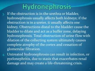
ANTENATAL HYDRONEPHROSIS. DR. A labdulaali ibrahim RIYADH MILITARY HOSPITAL. Introduction. Prenatal ultrasonography surveillance detects a significant fetal anomaly in 1% of pregnancies, of which 20-30% of cases are genitourinary in origin, and 50% manifest as hydronephrosis .

ANTENATAL HYDRONEPHROSIS
E N D
Presentation Transcript
ANTENATAL HYDRONEPHROSIS DR. Alabdulaaliibrahim RIYADH MILITARY HOSPITAL
Introduction • Prenatal ultrasonography surveillance detects a significant fetal anomaly in 1% of pregnancies, • of which 20-30% of cases are genitourinary in origin, and 50% manifest as hydronephrosis.
Introduction • At 20 weeks' gestation, the fetus is larger, and an anomaly is easier to detect. • Also by that time the amniotic fluid (AF) shifts from where most of it is placental transudate to where it becomes predominantly a product of fetal urine.
Frequency • The reported incidence of antenatal hydronephrosis ranges from 0.6 to 4.5 percent of pregnancies.
Frequency • Race • No known studies report the incidence of antenatal hydronephrosis related to race. • Sex • Hydronephrosis occurs approximately twice as often in males than in females. • Age • Studies have uniformly shown that timing of hydronephrosis is important. Early onset of hydronephrosis in fetal development is directly related to prognosis.
Mortality/Morbidity • Determination of mortality is difficult with antenatal hydronephrosis because of the significant incidence of stillbirths, terminated pregnancies, and missed diagnoses, all of which lead to underestimation of true mortality rates. • However, most research suggests that morbidity and mortality are directly related to the underlying etiology of hydronephrosis and the effect that the lesion has on the laterality, degree, and timing of hydronephrosis and resultant oligohydramnios.
Mortality/Morbidity • The survival rate with unilateral renal obstruction approaches 100%, with only 15-25% of patients requiring surgery at 4 years' follow-up.
Mortality/Morbidity • In the presence of a bilateral obstructive process, oligohydramnios is the best predictor of an adverse outcome. • The timing of oligohydramnios is an important determinant of fetal outcome. • The earlier a lesion develops, the more likely it is to have an effect on the fetal kidney, lungs, and overall outcome of the fetus.
History The finding of antenatal hydronephrosis should prompt a series of inquires regarding: Bladder cycling, Other anomalies, Prior pregnancy complications, Family history of urologic disease. • Onset, • Fetal sex, • Oligohydramnios, • Laterality, • Severity of hydronephrosis,
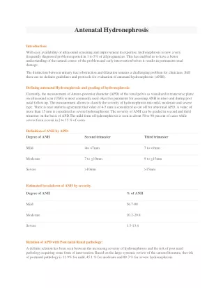
DEFINITION AND GRADING • Several studies have evaluated the threshold of fetal hydronephrosis resulting in clinically significant neonatal disease. • There are persistent postnatal renal abnormalities when the anteroposterior diameter (APD) of the fetal renal pelvis measures >5mm at <20 weeks,>8mm at 20±30 weeks and >10mm at >30 weeks of Gestation.
Society of Fetal Urology grading system for hydronephrosis Grade 1: renal pelvis is only visualized. Grade 2: renal pelvis as well as a few, but not all, calyces are visualized. Grade 3: virtually all calyces are visualized. Grade 4: similar to Grade 3 but, when compared to the normal centralateral kidney, there is parenchymalthinning.
Causes • Antenatal hydronephrosis without associated urinary tract anomaly is the etiology in the vast majority of infants with hydronephrosis (79-84%) and has been termed isolated antenatal hydronephrosis (IAHN). • IAHN is believed to be caused by a physiologic dilatation of the developing ureter.
The goal of evaluation is to differentiate benign physiologic dilation from significant obstructive disease or reflux.
Causes of antenatal hydronephrosis and their relative frequency • Transient 48 percent • Physiologic 15 percent • UPJ obstruction 11 percent • VUR 9 percent • Megaureter 4 percent • Multicystic dysplastic kidney 2 percent • Ureterocele 2 percent • Posterior urethral valves 1 percent Woodward M; Frank D; BJU Int 2002 Jan;89(2):149-56
Causes of antenatal hydronephrosis and their relative frequency • Less common causes included • Ectopic ureter, • Prune belly, • Urachal cyst, • Duplex collecting system, • Urethral atresia.
TREATMENT OF ANTENATAL HYDRONEPHROSIS • Intervention for a fetus with antenatal hydronephrosis is controversial. • The main considerations in evaluating a fetus with antenatal hydronephrosis are gestational age, laterality of the lesion, the presence of unfavorable prognostic factors, volume of amniotic fluid, and overall fetal well-being.
TREATMENT OF ANTENATAL HYDRONEPHROSIS • Predictors of poor prognosis: • Oligohydramnios is the most significant indicator of poor fetal outcome, • Presence of multiple fetal anomalies or chromosomal anomalies, • Bilateral lesions, • Renal cortical cysts and echogenic parenchyma, • Elevated levels of urinary electrolytes on vesicocentesis, • Reduced lung volume.
TREATMENT OF ANTENATAL HYDRONEPHROSIS • Significant unilateral hydronephrosis does not require prenatal intervention; • However, it should be evaluated in the postnatal period with follow-up renal ultrasonography (if needed), voiding cystourethrography, and diuretic renography.
TREATMENT OF ANTENATAL HYDRONEPHROSIS • Bilateral hydronephrosis without bladder distension is more significant and should be monitored prenatally with serial ultrasonographic examinations to monitor for bladder distension and development of oligohydramnios. • Postnatal evaluation should be performed with follow-up renal ultrasonography, voiding cystourethrography, and diuretic renography.
TREATMENT OF ANTENATAL HYDRONEPHROSIS • In the presence of oligohydramnios, evaluation for the presence of unfavorable prognostic factors with karyotype analysis and vesicocentesis is warranted. • Early delivery after giving steroid for lung maturation. • Fetuses with findings consistent with a poor outcome are generally not good candidates for prenatal intervention or ?? Pregnancy termination.
TREATMENT OF ANTENATAL HYDRONEPHROSIS • At present, fetal intervention for obstructive uropathy is indicated when the life of the neonate is at risk. • This is when oligohydramnios is present in the setting of presumed bladder outlet obstruction. • It is also essential that no other life-threatening conditions, particularly cardiovascular or neurologic, exist. • It is essential that there is a reasonable chance that the fetus will benefit from in utero decompression of the bladder.
TREATMENT OF ANTENATAL HYDRONEPHROSIS • Diagram showing technique of fetal vesicoamniotic shunt placement
THE GOAL • Is to identify patients with significant renal and urinary tract abnormalities while avoiding unnecessary testing in patients with physiologic or clinically insignificant hydronephrosis. • Evaluation includes physical examination and the use of radiologic studies to detect renal and urinary tract abnormalities including obstructive uropathy or vesicoureteral reflux (VUR).
Currently, there is not a single test or finding that accurately differentiates infants with significant disease from those who are normal or have insignificant findings.
PHYSICAL EXAMINATION • General apperance: • Syndrominc, • Outer ear anomaly. • Abdominal exam: • Mass that could represent an enlarged kidney due to obstructive uropathy or multicystic dysplastic kidney (MCDK). • A palpable bladder in a male infant, especially after voiding, may suggest posterior urethral valves. • Prune belly syndrome (deficient abdominal wall musculature and undescended testes). • Back exam.
Ultrasonography • The timing of the study depends upon the severity of the antenatal hydronephrosis. • In general, examination should be avoided in the first two days after birth because hydronephrosis may not be detected because of extracellular fluid shifts that will underestimate the degree of hydronephrosis. • Infants with bilateral hydronephrosis and those with a severe hydronephrotic solitary kidney require urgent evaluation on the first postnatal day because of the increased likelihood of significant disease.
Voiding cystourethogram • It is performed to detect VUR and in boys, to evaluate the posterior urethra.
Diuretic renography • Is used to diagnose urinary tract obstruction in infants with persistent hydronephrosis, usually ordered after a VCUG has demonstrated no vesicoureteral reflux. • It measures the drainage time from the renal pelvis and assesses total and each individual kidney's renal function. • The preferred radioisotope is technetium-99m-mercaptoacetyltriglycine (Tc99mMAG3)
MANAGEMENT APPROACH • Generally based upon the confirmation of persistent postnatal hydronephrosis and the following two predicative factors. • Bilateral involvement • Severe hydronephrosis— RPD >15 mm during the third trimester.
BILATERAL HYDRONEPHROSIS • Ultrasonography on the first postnatal day if severe or involving a solitary kidney. • If the postnatal ultrasound demonstrates persistent hydronephrosis, a VCUG should be performed to rule out ureterocoele or posterior urethral valves (PUV) in a male infant. • Infants with mild or moderate hydronephrosis can be evaluated after 7 days of life.
SEVERE UNILATERAL HYDRONEPHROSIS • In newborns with severe antenatal unilateral hydronephrosis (renal pelvic diameter >15 mm in the third trimester), • Ultrasonography should be performed after the infant returns to birth weight (after 48 hours of age and within the first two weeks of life).
MODERATE AND MILD UNILATERAL HYDRONEPHROSIS • In newborns with less severe antenatal unilateral hydronephrosis (renal pelvic diameter <15 mm during third trimester), ultrasonography can be performed after they reach seven days of age. • Antibiotic prophylaxis should therefore be considered until the VCUG has been performed and either the diagnosis of VUR has made or eliminated.
Antibiotic prophylaxis • Higher rates of urinary tract infections have been reported in children with prenatally diagnosed hydronephrosis compare to the general pediatric population. • The risk of infection rises if there is an underlying urologic abnormality, such as VUR or obstructive uropathy, and is greater in girls compared to boys. • As a result, in infants with severe hydronephrosis who are at greater risk for an underlying urologic abnormality, antibiotic prophylaxis (amoxicillin, 12 to 25 mg/kg PO daily) is started after delivery until the diagnosis of VUR or obstructive uropathy is excluded.
REFERENCES • Overview of antenatal hydronephrosis,Laurence Baskin & TulinOzcan, updated on June 16,2008. • Postnatal management of antenatal hydronephrosis, Laurence Baskin, updated on June 24,2008. • eMedicine: Antenatal Hydronephrosis, Dennis B Liu, John D Edmondson, Updated on Dec 11, 2008. • Campbell-Walsh Urology 9th edition.