RESPIRATION
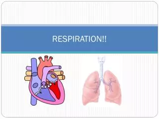
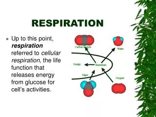
RESPIRATION. Gas exchange is the uptake of molecular oxygen (O 2 ) from the environment and the discharge of carbon dioxide (CO 2 ) to the environment. It is distinct from, but linked to, the production of ATP in cellular respiration.

RESPIRATION
E N D
Presentation Transcript
Gas exchange is the uptake of molecular oxygen (O2) from the environment and the discharge of carbon dioxide (CO2) to the environment. It is distinct from, but linked to, the production of ATP in cellular respiration. The oxygen source, the respiratory medium, is air for terrestrial animals and water for aquatic animals. The atmosphere is about 21% O2 (by volume). Dissolved oxygen levels in lakes, oceans, and other bodies of water vary considerably, but they are always much less than an equivalent volume of air. Gas exchange, in concert with the circulatory system, provide the oxygen necessary for aerobic cellular respiration and removes the waste product, carbon dioxide.
Gas exchange with the environment at the respiratory surface. Movements of CO2 and O2 across surface occurs entirely by diffusion. The rate of diffusion is proportional to the surface area. Therefore, respiratory surfaces tend to be thin and have large areas, maximizing the rate of gas exchange. In addition, the respiratory surfaces are moist to maintain the cell membranes and thus gases must first dissolve in water. The structure of a respiratory surface depends mainly on the size of the organism, whether it lives in water or on land, and by its metabolic demands. An endotherm has a larger area of respiratory surface than a similar-sized ectotherm.
Gas exchange occurs over the entire surface area of protists and other unicellular organisms. Similarly, for some relatively simple animals, such as sponges, cnidarians, and flatworms, the plasma membrane of every cell in the body is close enough to the outside environment for gases to diffuse in and out. In most animals, the bulk of the body lacks direct access to the respiratory medium. Some animals, such as earthworms and some amphibians, use the entire outer skin as a respiratory organ. Just below the moist skin is a dense net of capillaries. However, because the respiratory surface must be moist, their possible habitats are limited to water or damp places.
Animals that use their moist skin as their only respiratory organ are usually small and are either long and thin or flat in shape, with a high ratio of surface area to volume. Most other animals, general body surface lacks sufficient area to exchange gases for the entire body. How do they solve the problem? A respiratory organ that is extensively folded or branched, enlarging the surface area for gas exchange. Gills, tracheae, and lungs are the three most common respiratory organs. The total surface area of gills is often much greater than that of the rest of the body.
Water has both advantages and disadvantages as a respiratory medium. There is no problem keeping the cell membranes of the respiratory surface moist. However, O2 concentrations in water are low, especially in warmer and saltier environments. Thus, gills must be very effective to obtain enough oxygen.
Ventilation, increases the flow of the respiratory medium over the respiratory surface, ensures that there is a strong diffusion gradient between the gill surface and the environment. Without ventilation, a region of low O2 and high CO2 concentrations can form around the gill as it exchanges gas with the environment. Fish gills are ventilated by a current of water that enters the mouth, passes through slits in the pharynx, flows over the gills, and exits the body. Operculum moves the water. Water is dense and contains little oxygen per unit volume, so fishes expend considerable energy in ventilating their gills. Gas exchange at the gill surface is enhanced by the opposing flows of water and blood at the gills. Countercurrent flow
This flow pattern is countercurrent exchange. As blood moves anteriorly in a gill capillary, it becomes more and more loaded with oxygen, but it simultaneously encounters water with even higher oxygen concentrations because it is just beginning its passage over the gills. All along the gill capillary, there is a diffusion gradient favoring the transfer of oxygen from water to blood.
Gills are generally unsuited for an animal living on land. Why? An expansive surface of wet membrane exposed to air would lose too much water by evaporation. Gills would collapse as their fine filaments, no longer supported by water, would cling together, reducing surface area for exchange. Most terrestrial animals have their respiratory surfaces within the body, opening to the atmosphere through narrow tubes.
Advantages of air over water as a medium. Much higher concentration of oxygen. O2 and CO2 diffuse much faster in air than in water, so respiratory surfaces exposed to air do not have to be ventilated as thoroughly as gills. When a terrestrial animal does ventilate, less energy is needed because air is far lighter and much easier to pump than water and much less volume needs to be breathed to obtain an equal amount of O2. Air does have problems as a respiratory medium. The respiratory surface, which must be large and moist, continuously loses water to the air by evaporation. This problem is greater reduced by a respiratory surface found inside the body
Amphibians have relatively small lungs. Problem? They rely heavily on diffusion across their moist skin, for gas exchange. In contrast, most reptiles and all birds and mammals rely entirely on lungs for gas exchange.
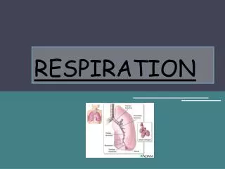
Air enters through the nostrils. What happens there? The nasal cavity leads to the pharynx, and when the glottis is open, air enters the larynx, the upper part of the respiratory tract. In most mammals, the larynx is adapted as a voicebox in which vibrations of a pair of vocal cords produce sounds Next the trachea, or windpipe, rings of cartilage. Two bronchi, within the lung, each bronchus branches into finer and finer tubes, called bronchioles. The epithelium lining is covered by cilia and a thin film of mucus. The bronchioles dead-end as a cluster of air sacs called alveoli. Gas exchange occurs across the thin epithelium of the lung’s millions of alveoli with a total surface area of about 100 m2 in humans. Oxygen dissolves in the moist film and rapidly diffuses across the epithelium into a web of capillaries that surrounds each alveolus.
The process of breathing, the alternate inhalation and exhalation of air, ventilates lungs. A frog ventilates its lungs by positive pressure breathing. During a breathing cycle, muscles lower the floor of the oral cavity, enlarging it and drawing in air through the nostrils. With the nostrils and mouth closed, the floor of the oral cavity rises and air is forced down the trachea. Elastic recoil of the lungs, together with compression of the muscular body wall, forces air back out of the lungs during exhalation.
In contrast, mammals ventilate their lungs by negative pressure breathing. This works like a suction pump, pulling air instead of pushing it into the lungs. Muscle action changes the volume of the rib cage and the chest cavity,and the lungsfollow suit.
Lung volume increases as a result of contraction of the rib muscles and diaphram. Inhilation, Inspiration. Contraction of the rib muscles expands the rib cage by pulling the ribs upward and the breastbone outward. At the same time, the diaphram contracts and descends. These changes increase the lung volume, and as a result, air pressure within the alveoli becomes lower than atmospheric pressure. Because air flows from higher pressure to lower pressure, air rushes into the respiratory system. During exhalation, the opposite occurs. Usually about a 10% change Actions of the rib muscles and diaphragm accounts for changes in lung volume during shallow breathing, when a mammal is at rest. During vigorous exercise, other muscles of the neck, back, and chest further increase ventilation volume by raising the rib cage even more.
The volume of air an animal inhales and exhales with each breath is called tidal volume. 1. Anatomical dead space a. 150 ml within air passages. Never goes to the alveoli. b. Explains the drop in partial pressure of oxygen from out to inside of alveoli. Makes alveoli much less efficient than gills. c.. This 2 way flow reduces water loss for land animals It averages about 500 mL in resting humans. Can be increased to 3,000 mL during exercise The maximum tidal volume during forced breathing is the vital capacity, which is about 3.4 L and 4.8 L for college-age females and males, respectively. The lungs hold more air than the vital capacity, but some air remains in the lungs, the residual volume, because the alveoli do not completely collapse.
Respiratory rate: number of breaths per unit time(10-15) Minute respiratory volume (MRV) 1. Tidal volume x respiratory rate per minute. Air entering and leaving lung per minute 2. Normally 5 liters/minute. Can be as high as 130 liters/minute-exercise. 3. MRV extremely high a. CO2 removed from blood by ventilation faster than its produced by tissues-raises pH-basic b. Hyperventilation-basic pH doesn’t trigger response c. Not O2 level increase-takes time to stimulate breath response 4. MRV unusually low a. Elevated P CO2 level-lower pH triggers respiratory response b. Hypoventilation(holding your breath)
Our breathing control centers are located in two brain regions, the medulla oblongata and the pons. • Aided by the control center in the pons, the medulla’s center sets basic breathing rhythm, triggering contraction of the diaphragm and rib muscles. • A negative-feedback mechanism via stretch receptors prevents our lungs from overexpanding by inhibiting the breathing center in the medulla.
The medulla’s control center monitors the CO2 level of the blood and regulated breathing activity appropriately. Its main cues about CO2 concentration come from slight changes in the pH of the blood and cerebrospinal fluid bathing the brain Carbon dioxide reacts with water to form carbonic acid, which lowers the pH. When the control center registers a slight drop in pH, it increases the depth and rate of breathing, and the excess CO2 is eliminated in exhaled air. Oxygen concentrations in the blood usually have little effect of the breathing control centers. When the O2 level is severely depressed - at high altitudes, for example, O2 sensors in the aorta and carotid arteries in the neck send alarm signals to the breathing control centers, which respond by increasing breathing rate. However, deep, rapid breathing purges the blood of so much CO2 that the breathing center temporarily ceases to send impulses to the rib muscles and diaphragm.
The respiratory center responds to a variety of nervous and chemical signals and adjusts the rate and depth of breathing to meet the changing demands of the body. Control is only effective if it is coordinated with control of the circulatory system, so that there is a good match between lung ventilation and the amount of blood flowing through alveolar capillaries. For example, during exercise, cardiac output is matched to the increased breathing rate, which enhances O2 uptake and CO2 removal as blood flows through the lungs.
For a gas, whether present in air or dissolved in water, diffusion depends on differences in a quantity called partial pressure, the contribution of a particular gas to the overall total. At sea level, the atmosphere exerts a total pressure of 760 mm Hg. Since the atmosphere is 21% oxygen (by volume), the partial pressure of oxygen (abbreviated PO2) is 0.21 x 760, or about 160 mm Hg. The partial pressure of CO2 is only 0.23 mm Hg. A gas will always diffuse from a region of higher partial pressure to a region of lower partial pressure.
Blood arriving at the lungs via the pulmonary arteries has a lower PO2 and a higher PCO2 than the air in the alveoli. • As blood enters the alveolar capillaries, CO2 diffuses from blood to the air within the alveoli, and oxygen in the alveolar air dissolves in the fluid that coats the epithelium and diffuses across the surface into the blood. • By the time blood leaves the lungs in the pulmonary veins, its PO2 have been raised and its PCO2 has been lowered.
In the tissue capillaries, gradients of partial pressure favor the diffusion of oxygen out of the blood and carbon dioxide into the blood. • Cellular respiration removes oxygen from and adds carbon dioxide to the interstitial fluid by diffusion, and from the mitochondria in nearby cells. • After the blood unloads oxygen and loads carbon dioxide, it is returned to the heart and pumped to the lungs again, where it exchanges gases with air in the alveoli.
The low solubility of oxygen in water is a fundamental problem for animals that rely on the circulatory systems for oxygen delivery. For example, a person exercising consumes almost 2 L of O2 per minute, but at normal body temperature and air pressure, only 4.5 mL of O2 can dissolve in a liter of blood in the lungs. If 80% of the dissolved O2 were delivered to the tissues (an unrealistically high percentage), the heart would need to pump 500 L of blood per minute - a ton every 2 minutes. In fact, most animals transport most of the O2 bound to special proteins called respiratory pigments instead of dissolved in solution. Found in blood. Special cell. The presence of respiratory pigments increases the amount of oxygen in the blood to about 200 mL of O2 per liter of blood. For our exercising individual, the cardiac output would need to be a manageable 20-25 L of blood per minute to meet the oxygen demands of the systemic system. Respiratory pigments transport gases and help buffer the blood
A diversity of respiratory pigments have evolved. • Hemocyanin, found in arthropods and many mollusks, has copper as its oxygen-binding component, coloring the blood bluish. • The respiratory pigment of almost all vertebrates is the protein hemoglobin, in red blood cells.
Carbon dioxide from respiring cells diffuses into the blood plasma and then into red blood cells, where some is converted to bicarbonate, assisted by the enzyme carbonic anhydrase. • At the lungs, the equilibrium shifts in favor of conversion of bicarbonate to CO2.
Hemoglobin must bind oxygen reversibly, loading oxygen at the lungs or gills and unloading it in other parts of the body. Loading and unloading depends on cooperation among the subunits of the hemoglobin molecule. The binding of O2 to one subunit induces the remaining subunits to change their shape slightly such that their affinity for oxygen increases. When one subunit releases O2, the other three quickly follow suit as a conformational change lowers their affinity for oxygen.
Cooperative oxygen binding and release is evident in the dissociation curve for hemoglobin. Where the dissociation curve has a steep slope, even a slight change in PO2 causes hemoglobin to load or unload a substantial amount of O2. This steep part corresponds to the range of partial pressures found in body tissues. Hemoglobin can release an O2 reserve to tissues with high metabolism.
Like all proteins, hemoglobin’s conformation is sensitive to a variety of factors. • A drop in pH lowers the affinity of hemoglobin for O2, an effectcalled the Bohr shift. • Because CO2 reacts with water to form carbonic acid, an active tissue will lower the pH of its surroundingsand induce hemoglobinto release more oxygen because it has a lower affinity for oxygen. • Dissociation curve shifted to right. • This occurs in red blood cells • Hemoglobin releases oxygen more readily
In addition to oxygen transport, hemoglobin also helps transport carbon dioxide and assists in buffering blood pH. About 7% of the CO2 released by respiring cells is transported in solution. Another 23% binds to amino groups of hemoglobin. About 70% is transported as bicarbonate ions.
When an air-breathing animal swims underwater, it lacks access to the normal respiratory medium. Most humans can only hold their breath for 2 to 3 minutes and swim to depths of 20 m or so. However, a variety of seals, sea turtles, and whales can stay submerged for much longer times and reach much greater depths. How? 7. Deep-diving air-breathers
These deep-divers, such as the Weddell seal, is an ability to store large amounts of O2 in the tissues. Compared to a human, a seal can store about twice as much O2 per kilogram of body weight, mostly in the blood(oxyhemoglobin) and muscles(myoglobin). About 36% of our total O2 is in our lungs and 51% in our blood. Weddell seal holds only about 5% of its O2 in its small lungs and stockpiles 70% in the blood. Several adaptations create these physiological differences between the seal and other deep-divers in comparison to humans. First, the seal has about twice the volume of blood per kilogram of body weight as a human. Second, the seal can store a large quantity of oxygenated blood in its huge spleen, releasing this blood after the dive begins. Third, diving mammals have a high concentration of an oxygen-storing protein called myoglobin in their muscles. This enables a Weddell seal to store about 25% of its O2 in muscle, compared to only 13% in humans.
Diving vertebrates start a dive with a relatively large O2 stockpile, but they also have adaptations that conserve O2. They swim with little muscular effort and often use buoyancy changes to glide passively upward or downward. Their heart rate and O2 consumption rate decreases during the dive and most blood is routed to the brain, spinal cord, eyes, adrenal glands, and placenta (in pregnant seals). Blood supply is restricted or even shut off to the muscles, and the muscles can continue to derive ATP from fermentation after their internal O2 stores are depleted.
Ventilation is much more complex in birds than in mammals. Besides lungs, birds have eight or nine air sacs that do not function directly in gas exchange, but act as bellows that keep air flowing through the lungs.
The entire system - lungs and air sacs - is ventilated when the bird breathes. Air flows through the interconnected system in a circuit that passes through the lungs in one direction only, regardless of whether the bird is inhaling or exhaling. Instead of alveoli, which are dead ends, the sites of gas exchange in bird lungs are tiny channels called parabronchi, through which air flows in one direction.
This system completely exchanges the air in the lungs with every breath. Therefore, the maximum lung oxygen concentrations are higher in birds than in mammals. Partly because of this efficiency advantage, birds perform much better than mammals at high altitude. For example, while human mountaineers experience tremendous difficulty obtaining oxygen when climbing the Earth’s highest peaks, several species of birds easily fly over the same mountains during migration.