Download

1 / 32
360 likes | 700 Vues
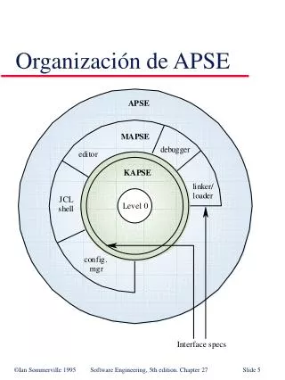
Basic Biomechanics , (5th edition) by Susan J. Hall, Ph.D. Chapter 8 The Biomechanics of the Human Lower Extremity. Structure of the Hip. What is the hip joint ?. a ball and socket joint where the head of the femur articulates with the concave acetabulum

E N D
Basic Biomechanics, (5th edition)by Susan J. Hall, Ph.D. Chapter 8 The Biomechanics of the Human Lower Extremity
Structure of the Hip What is the hip joint? • a ball and socket joint • where the head of the femur articulates with the concave acetabulum • a more stable joint than the shoulder because of bone structure and the number and strength of the muscles and ligaments crossing the joint
Iliofemoral (Y) ligament Pubofemoral ligament Femur Iliofemoral ligament Ischium Ischium Femur Ischiofemoral ligament Anterior view Posterior view Structure of the Hip The integrity of the hip is enhanced by the strong ligaments crossing the joint.
Sacrum Ilium Acetabulum Femoral head Pubis Ischium Femur Structure of the Hip The pelvic girdle includes the two ilia and the sacrum. It can be rotated forward, backward, and laterally to optimize positioning of the hip.
Movements at the Hip What movements of the femur are facilitated by pelvic tilt? Pelvic tilt directionFemoral movement posterior flexion anterior extension lateral (to opposite abduction side)
Movements at the Hip What muscles contribute to flexion at the hip? • iliacus • psoas major • assisted by: • pectineus • rectus femoris • sartorius • tensor fascia latae
Movements at the Hip What muscles contribute to extension at the glenohumeral joint? • gluteus maximus • hamstrings • biceps femoris • semimembranosus • semitendinosus
Movements at the Hip What muscles contribute to abduction at the glenohumeral joint? • gluteus medius • assisted by: • gulteus minimus
Movements at the Hip What muscles contribute to adduction at the glenohumeral joint? • adductor magnus • adductor longus • adductor brevis • assisted by: • gracilis
Structure of the Knee What is the tibiofemoral joint? • dual condyloid articulations between the medial and lateral condyles of the tibia and the femur; composing the main hinge joint of the knee • considered to be the knee joint
Femur Patella Tibia Fibula Structure of the Knee Bony structure of the tibiofemoral joint.
Structure of the Knee What is the patellofemoral joint? • articulation between the patella and the femur • (the patella improves the mechanical advantage of the knee extensors by as much as 50%)
Structure of the Knee What are the menisci? • cartilaginous discs located between the tibial and femoral condyles • structures that distribute the load at the knee over a large surface area and also help absorb shock
Transverse ligament Lateral meniscus Medial meniscus Anterior cruciate ligament Posterior cruciate ligament Superior view Structure of the Knee The menisci of the knee.
Structure of the Knee What major ligaments cross the knee? • collateral ligaments - cross the medial and lateral aspects of the knee • cruciate ligaments - cross each other in connecting the anterior and posterior aspects of the knee
Movements at the Knee What muscles contribute to flexion at the knee? • popliteus - “unlocks” the fully extended knee by laterally rotating the femur with respect to the tibia to allow flexion to proceed
Movements at the Knee What muscles contribute to flexion at the knee? • hamstrings • assisted by: • gracilis • sartorius • popliteus • gastrocnemius
Movements at the Hip What muscles contribute to extension at the hip? • quadriceps: • rectus femoris • vastus lateralis • vastus medialis • vastus intermedius
Structure of the Ankle What is the tibiotalar joint? • hinge joint where the convex surface of the superior talus articulates with the concave surface of the distal tibia • considered to be the ankle joint
Structure of the Ankle What is the distal tibiofibular joint? (a syndesmosis where dense, fibrous tissue binds the distal tibia and fibula together)
Fibula Tibia Talus Calcaneus Posterior view Structure of the Ankle The bony structure of the ankle.
Movements at the Ankle What muscles contribute to dorsiflexion at the ankle? • tibialis anterior • extensor digitorum longus • peroneus tertius • assisted by: • extensor hallucis longus
Movements at the Ankle What muscles contribute to plantar flexion at the ankle? • gastrocnemius • soleus • assisted by: • tibialis posterior, plantaris, peroneus longus, flexor hallucis longus, peroneus brevis, flexor digitorum longus
Structure of the Foot What is the subtalar joint? (the anterior and posterior facets of the talus articulate with the sustencalculum tali on the superior calcaneus)
Structure of the Foot What are the tarsometatarsal and intermetatarsal joints? • nonaxial joints that permit only gliding movements • enable the foot to function as a semirigid unit and to adapt flexibly to uneven surfaces during weight bearing
Structure of the Foot What are the metatarsophalangeal and interphalangeal joints? • condyloid and hinge joints, respectively • the toes function to smooth the weight shift to the opposite foot during walking and help maintain stability during weight bearing by pressing against the ground when necessary
Structure of the Foot What are the plantar arches? • the medial and lateral longitudinal arches stretch form the calcaneus to the metatarsals and tarsals • the transverse arch is formed by the bases of the metatarsal bones
Structure of the Foot What are the plantar fascia? • thick bands of fascia that cover the plantar aspects of the foot • During weight bearing, mechanical energy is stored in the stretched ligaments, tendons, and plantar fascia of the foot. This energy is released to assist with push-off of the foot from the surface.
Lateral view Plantar fascia Plantar view Structure of the Foot The plantar fascia.
Movements of the Foot What muscles are responsible for toe flexion and extension? • flexion - flexor digitorum longus, flexor digitorum brevis, quadratus plantae, lumbricals, interossei • extension - extensor hallucis longus, extensor digitorum longus, extensor digitorum brevis
Movements of the Foot What muscles are responsible for inversion and eversion? • inversion - tibialis posterior, tibialis anterior • eversion - peroneus longus, peroneus brevis, assisted by peroneus tertius
Chapter 8 The Biomechanics of the Human Lower Extremity