Download
1 / 42
440 likes | 760 Vues
(Chapters 6-7 of KS). Ion transport across the cell membrane underlies cellular Homeostasis and electrical activity. 1.- the cell membrane. 2.- ion transport across membranes . 3.- ion channels structure and function . 4.- osmotic balance and ion channels .

E N D
(Chapters 6-7 of KS) Ion transport across the cell membrane underlies cellular Homeostasis and electrical activity 1.- the cell membrane 2.- ion transport across membranes 3.- ion channels structure and function 4.- osmotic balance and ion channels 5.- ion channels and the control of membrane potential Readings: 1.- Neher E, Sakmann B. "The patch clamp technique" Sci Am. 1992 Mar;266(3):44-51. 2.- Doyle et al, “The structure of the potassium channel: molecular basis of K+ conduction and selectivity” Science. 1998 Apr 3;280(5360):69-77.
Problems for these chapters: Describe, quantitatively, the series of electrical events that follow the opening of a single Na+ selective ion channel (10 pS) in the membrane of an isolated vesicle. The lipid vesicle has a diameter of 3 ~micrometers. The concentration of Na+ outside is 150 mM. The internal Na+ concentration is 5 mM. Determine the polarity, magnitude and time course of the changes. Five seconds after opening, the Na+ channel closes. Then, a K+ selective ion channel opens for five seconds (Koutside = 5 mM; Kinside = 150 mM). How much are the internal Na+ and K+ concentrations changed in each cycle. What will happen if this cycle is repeated several thousand times? Describe the changes in membrane potential, cell volume and water flow that occur after reducing the external Cl- concentration in the solution bathing a single frog muscle fiber (slide #39)
Keywords: Bilayer size, properties, membrane capacitance Ion channel structure, single ion channel currents Faraday, membrane potential, charge Nernst equation Donnan equilibrium electrical equivalent circuits Permeability ratios Goldman equation Electrical Driving force
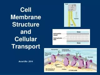
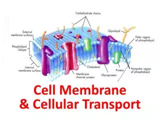
Ultrastructure of a typical animal cell The cell membrane contains many proteins including ion channels
Transmission electron micrograph of a cell membrane. The photograph shows two adjacent cells of the pancreas of a frog at a magnification of ×43,000. The inset is a high-magnification view (×216,000) of the plasma membranes of the cells. Note that each membrane includes two dense layers with an intermediate layer of lower density. The dense layers represent the interaction of the polar head groups of the phospholipids with the OsO4 used to stain the preparation. (From Porter KR, Bonneville MR: Fine Structure of Cells and Tissues, 4th ed., Philadelphia, Lea & Febiger, 1973.)
Structure of ion channels. Most ion channels consist of four to six subunits that are arranged like a rosette in the plane of the membrane. The channel can be made up of (1) identical, distinct subunits (homo-oligomer); (2) distinct subunits that are homologous but not identical (hetero-oligomer); or (3) repetitive subunit-like domains within a single polypeptide (pseudo-oligomer). In any case, these "subunits" surround the central pore of the ion channel. Note that each "subunit" is itself made up of several transmembrane segments.
Formation of an aqueous pore by an ion channel. The dielectric constant of water (ε= 80)is about 40-fold higher than the dielectric constant of the lipid bilayer(ε = 2).
Diffusion potential across a planar lipid bilayer containing a K+-selective channel
Patch clamp methods. (Data from Hamill OP, Marty A, Neher E, Sakmann B, Sigworth FJ: Improved patch-clamp techniques for high-resolution current recording from cells and cell-free membrane patches. Pflugers Arch 391:85-100, 1981.)
Three-dimensional image of the nicotinic acetylcholine receptor channel. (Data from Toyoshima C, Unwin N: Ion channel of acetylcholine receptor reconstructed from images of postsynaptic membranes. Nature 336:247-250, 1988.)
Subunit structure and membrane-folding models of voltage-gated channels. A, A voltage-gated Na+ channel is made up of a pseudo-oligomeric a subunit, as well as membrane-spanning b1 and b2 subunits. Note that the domains I through IV of the a subunit are homologous to a single subunit of a voltage-gated K+ channel (see C). B, A voltage-gated Ca2+ channel is made up of a pseudo-oligomeric a1 subunit, as well as an extracellular a2 subunit, a cytoplasmic b subunit, and membrane-spanning g and d subunits. Note that the domains I through IV of the a subunit are homologous to a single subunit of a voltage-gated K+ channel (see C).
A voltage-gated K+ channel is made up of four a subunits, as well as a cytoplasmic a subunit. (Data from Isom LL, De Jongh KS, Catterall WA: Auxiliary subunits of voltage-gated ion channels. Neuron 12:1183-1194, 1994.)
Some mutations of human Na+ channels. At least two genetic diseases are caused by mutations in the Na+ channel of human skeletal muscle. Hyperkalemic periodic paralysis can be caused by mutations in membrane-spanning segment S5 of domain II and S6 of domain IV. Paramyotonia congenita can be caused by mutations in membrane-spanning segment S3 of domain IV and S4 of domain IV. The disease can also be caused by mutations in the intracellular segment that links domains III and IV. (Data from Catterall WA: Cellular and molecular biology of voltage-gated sodium channels. Physiol Rev 72:S15-S48, 1992.)
Structure of the Streptomyces K+ channel (KcsA). A, KcsA is a homotetramer. Each monomer is represented in a different color and contains only two membrane-spanning elements, which is analogous to the S5-P-S6 portion of Shaker-type K+ channels. B, This view more clearly shows the P region, which is very similar to the P region of the Shaker K+ channel. The P region appears to form the selectivity filter of the channel. C, This is a cut-away view of the pore that shows three K+ ions. The top two K+ ions are bound in a tight cage that is formed by the peptide backbones of the P regions of each of the four channel subunits. (Data from Doyle DA, Morais Cabral J, Pfuetzner RA, et al: The structure of the potassium channel: Molecular basis of K+ conduction and selectivity. Science 280:69-77, 1998.)
Ohm’s law An open ion channel follows Ohm’s law!
Voltage dependence of currents through single Cl channels in outside-out patches. A, The channel is a gamma-aminobutyric acid-A (GABAA) receptor channel, which is a Cl channel activated by GABA. Identical solutions, containing 145 mM Cl, were present on both sides of the patch. B, The magnitudes of the single-channel current transitions (y-axis) vary linearly with voltage (x-axis). (Data from Bormann J, Hamill OP, Sakmann B: Mechanism of anion permeation through channels gated by glycine and g-aminobutyric acid in mouse cultured spinal neurones. J Physiol (Lond) 385:243-286, 1987.)
Goldman-Hodgkin-Katz equation. Valid when the total membrane current equals zero; Im=IK+INa+ICl=0
Dependence of the resting membrane potential on [K+]o and on the PNa/Pk ratio, a. The blue line describes an instance in which there is no Na+ permeability (i.e., PNa/Pk = 0). The three orange curves describe the Vm predicted by the GHK Equation for a values greater than zero. The deviation of these orange curves from linearity is greater at low values of [K+]o, where the [K+]o is relatively larger.
DP=RTDOsm Gibbs-Donnan equilibrium. A semipermeable membrane separates two compartments that have rigid walls and equal volumes. The membrane is permeable to Na+, Cl, and water, but not to the macromolecule Y, which carries 150 negative charges.
Effect of urea on cell volume. Because the cell membrane is far more permeable to water than to urea, we will assume that, during the initial moments in steps 2 and 3, the cell membrane is permeable only to water. Later, during steps 4 and 5, we assume that the membrane is permeable to both water and urea.