Download
1 / 53
530 likes | 741 Vues
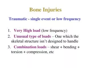
Stress Injuries of Bone in the Pediatric Athlete. Christopher Couture, MD Victory Sports Medicine May 8, 2012. Key Concepts. Skeletal Maturity Critical vs. Non-Critical Fractures. Skeletal Maturity. In skeletally mature, overuse of bone leads to stress fracture

E N D
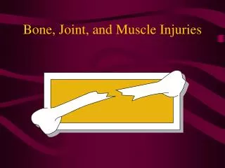
Stress Injuries of Bonein the Pediatric Athlete • Christopher Couture, MD • Victory Sports Medicine • May 8, 2012
Key Concepts • Skeletal Maturity • Critical vs. Non-Critical Fractures
Skeletal Maturity • In skeletally mature, overuse of bone leads to stress fracture • In bones with open physes, the injury occurs at the physis
Historical Features • Insidious onset activity-related pain that gradually worsens with time • Pain generally well localized • Pain relieved with rest, although over time can continue to occur at rest
Physical Exam • Localized tenderness • Rarely warmth, erythema, edema or (for fractures) palpable callus • Pain w/vibratory stimulus inconsistent • Special clinical tests specific to body area • i.e. “hop test” for femoral neck stress fx, “stork test” for pars intraarticularis stress fx
Radiographs • Very specific; not very sensitive • Usually negative early in course • 2/3 initially negative, only ½ of these become positive • Onset of pain can precede positive film by 2-3 months • Characteristic findings: • Skeletally immature: physeal widening, sclerosis or closure • Skeletally mature: periosteal thickening, radiolucent line formation
Triple-Phase/SPECT Scan • Very sensitive; not very specific • Can confirm diagnosis 2-8 days after sx onset • Can help to differentiate between other etiologies of symptoms (ex. medial tibial stress syndrome) • Characteristic findings: positive area of intense uptake on all three phases of scan
MRI • Best combination of sensitivity and specificity • Promising in grading progressive stages of severity • Can localize site of injury and differentiate from other entities such as tumor, osteomyelitis, bony infarct, etc.
Principles of Management • REDUCE ACTIVITY BELOW THRESHOLD FOR SYMPTOMS • Cessation or reduction of inciting sport • Immobilization • Non-weight-bearing • Orthotics (?) • Alternative training to maintain aerobic fitness, muscle tone, stamina • Other • Ice massage, NSAIDs, stretching/strengthening • “Invasive” treatment for displaced or nonhealing fractures
Physis (epiphyseal plate; pressure epiphysis): the segment of the bone that is responsible for lengthening • Apophysis (traction epiphysis): growth center at point where muscle can attach (tibial tubercle, calcaneus); contribute to bone shape but not length
Repetitive loading alters metaphyseal perfusion • Interferes with mineralization of hypertrophied chondrocytes • Hypertrophic zone continues to widen • Ischemia can lead to osseous necrosis & deformity • Can lead to localized, asymmetric growth or complete cessation of growth • ...although most stress injuries resolve without growth complications
Physeal injuries of Long Bone • Proximal humerus • Distal radius • Distal femur • Proximal tibia • Metatarsals • ANY long bone...
Proximal Humerus (Little Leaguer’s Shoulder) • Sequelae of repetitive traction/rotational forces across physis • Baseball pitchers, swimmers, gymnastics, volleyball • “Adolescent athlete’s shoulder”
Distal Radius • Gymnasts: Most commonly reported physeal stress injury • Radiographically widened physis especially on metaphyseal side • Usually recover with rest & do not experience abnormal growth
Apophyseal Injuries • Osgood-Schlatter • Sindig-Larsen-Johansson • Sever’s (calcaneal “apophysitis”) • Little Leaguer’s elbow (medial epicondyle) • Panner’s (osteochondrosis of capitellum) • Pelvic apophyses (ASIS, AIIS, iliac crest)
Reasons for Concern • Uninformed coaches and parents of children & adolescents • Increasing incidence of lower extremity stress injuries
Countermeasures • Individualized training & skill development • Delay progressions in periods of rapid growth • Variety - avoid repetition • Quality over volume • Periodic physical exam; directed studies • Physical conditioning • Trained personnel (certified athletic trainers) • Periodization • Appropriate assessment of physical maturity for expected demands
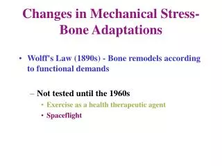
Wolff’s Law • Bone will remodel itself in response to mechanical stress (or more accurately, strain) • Sources of strain include muscle contraction & ground stresses • Bone responds by increasing its rate of remodeling • Resorption by osteoclasts precedes replacement by osteoblasts • Lag time – weaker structure during this period
How Does This Lead To Fracture? • If sufficient recovery time allowed, stronger bone results • If repeated application of stress, microdamage can accumulate • Compensatory remodeling is thought to be able to repair this microdamage, but if repetitive loading continues and remodeling cannot maintain integrity of bone, stress fractures develop • Influenced by number of repetitions, frequency of repetitions, amount of load
Risk Factors • ~ 60% of patients w/stress fracture have had prior stress fracture • Participation in sports involving running and jumping • Rapid increase in physical training program • Poor preparticipation physical condition • Female gender • Hormonal or menstrual disturbances • Decreased bone density • Decreased thickness of cortical bone • Nutritional deficiencies (including dieting) • Extremes of body size and composition • Running on irregular or angled surfaces • Inappropriate or worn-out footwear • Inadequate muscle strength • Poor flexibility • “Type A” behavior
Training Errors • Substantial, sudden increase in training intensity, duration or distance • Sudden change in training surface • Harder, uneven surfaces • Acquisition of new (“inadequate”) footwear • Or keeping old footwear too long (500 mi. rule)
Critical stress fractures require specific attention due to increased rates of nonunion • Include anterior tibia, medial malleolus, talus, navicular, 5th metatarsal, sesamoids • Usually treat nonweightbearing 6+ weeks • Noncritical stress fractures can be treated with a short period of immobilization or relative rest • Include medial tibia, fibula, 2nd–4th metatarsals • Usually return to play in 6-8 weeks
Medial Tibia • Posteromedial border of distal 1/3 of tibia • Association with high-arched feet or excessively flat feet • Walking boot • May begin gradual return to activity after tenderness disappears; usually full return to play in 8-12 weeks
Fibula • Minimal role in weight bearing • Stress fractures arise from muscle traction and torsional forces • Usually distal 1/3 • Treat with weight-bearing rest (4-6 weeks), perhaps with short period in walking boot
Metatarsals • Mets 1,3,4 and distal 2 usually uncomplicated and treated with relative rest; walking boot • Gradual return to sport program after tenderness resolved
Base of 2nd Metatarsal • Common among ballet dancers • Can be treated with weight-bearing rest but patients must refrain from training for 6 weeks
Calcaneus • Localized tenderness over medial or lateral aspects of calcaneus, usually at upper posterior margin • Treat: 6-8 weeks weight-bearing rest with soft heel cushion
Cuboid & Cuneiforms • Rare - usually considered non-critical • Treat: weight-bearing rest with short period in walking boot
Coronoid Process • From repeated impact in trapshooting • Presents with nonspecific aching in shoulder • Pain with palpation and resisted active adduction/forward flexion • Axillary view XR • Avoid shooting 6-8 weeks
Humerus • Proximal fractures, medial epicondyle fractures seen in throwers, gymnasts and pole vaulters • Midshaft fractures seen in throwers and workers doing heavy lifting • Pain with throwing or lifting may involve entire upper arm • Treat: rest 6-8 weeks
Radius • Uncommon location - reported in military personnel and tennis, volleyball and softball players, gymnasts and cheerleaders and football offensive linemen • Pain in shaft of radius with exertion; pain with weight-bearing on wrist or active wrist extension • Treat with 6 weeks immobilization
Ulna • Reported in softball pitchers, rodeo riders and volleyball players, racquet sports and football offensive linemen • Pain with underhand maneuvers • Treat with relative rest and modification of biomechanics if necessary
Ribs • First rib: Seen in baseball pitchers and basketball players • Pain with arm motion over supraclavicular area • Other ribs: Seen in softball players, golfers and rowers • Pain with rotation of the trunk • Noncritical: treat with relative rest and gradual return to activity
Spondylolysis • Pars intraarticularis stress fractures • Common in gymnasts, cheerleaders and weightlifters; L4-5 most common levels • Usually involved in repetitive extension load activity • Ultimately complains of significant back spasms (often coinciding with growth spurt) – often misdiagnosed as lumbar strain • Pain with one- or two-leg standing extension tests; gait abnormality 2ndary to hamstrings tightness • Plain XR usually negative; obliques - “collar on the Scottie dog” • Bone scan helpful; SPECT scan or MRI more sensitive • Treatment: controversial – rest +/- corset brace (thoracic lumbosacral orthosis); often requires 3-6 months
Spondylolisthesis • Bilateral pars defects allowing slippage of vertebral bodies • Graded depending on amount of slippage • Lateral view XR • High-grade may have radiculopathy • Treatment: Grade 1 or older adolescents with Grade 2 - intensive treatment similar to spondylolysis • Surgical stabilization if progressive, persistent pain or radiculopathy, younger children with Grade 2, all Grade 3-4 (L4-S1 fusion)
Femoral Neck • Seen in runners and dancers, especially older athletes • Critical - complications can be severe including AVN, nonunion, deformities and displacement • Present with pain in groin, anterior thigh or knee; aching pain usually precipitated by weight-bearing activity and brought about by significant change in training • Differentiate compression from traction lesion • Treatment: often surgery, especially for all tension lesions; compression lesions can be treated conservatively if mild but often requires bed rest (non-weight-bearing mandatory)
Anterior Cortex of Tibia • Prone to delayed union, nonunion and progression to complete fracture • Vulnerable to nonunion b/c poor vascularity and increased mechanical tension • “Dreaded black line” • Treatment programs include prolonged rest (4-6 months), bone stimulators/TENS; consideration of IM nailing immediately
Medial Malleolus of Tibia • Usually present with weeks of mild discomfort punctuated by acute episode • Excessive ankle pronation with accomp. tibial rotation distribute excessive forces to med mall • Nondisplaced fractures can be treated conservatively with pneumatic leg brace for 6 weeks • Displaced fractures or nonunions require surgery
Tarsal Navicular • Central 1/3: relative avascularity, shear forces in the region • Mild symptoms; vague dorsal pain in midfoot • Critical exam: pain to palpation in “N” spot • Advanced imaging should be pursued early if suspected • 6 weeks non-weight-bearing immobilization; if still tender, another 2 weeks; use physical therapy • If no response or displaced, surgery (screw fixation +/- bone grafting)
Talus • Usually involve lateral body; neck fractures rare • Often present with prolonged pain following ankle sprain despite adequate rehab • Excessive subtalar pronation allows lateral impingement of the lateral process of the calcaneus on the posterolateral corner of the talus • Treat: non-weight-bearing x 6 weeks • Nonunion: surgical excision of lateral process of talus
Proximal 5th Metatarsal • Tuberosity avulsion fracture • Usually result of acute inversion injury • Uncomplicated; treated with brief immobilization for pain relief followed by progressive activity • Jones fracture • Occurs at junction of metaphysis and diaphysis • Acute vs. chronic • Chronic: critical and prone to nonunion • Treated with 6-10 weeks of non-weight-bearing rest • Failure to heal or displaced: fixation screw (early?) • Diaphyseal stress fracture • As for chronic Jones fracture
Sesamoids • Medial and lateral sesamoids at 1st MTP act to increase mechanical advantage of FHB tendon and absorb weight-bearing stress on medial forefoot • Medial sesamoid more commonly affected • Usually requires advanced imaging; need to differentiate bipartite sesamoid from true fracture • Critical: prone to non-union • Treat: non-weight-bearing rest for 6 weeks • If nonunion or splintered, surgical excision