Download
1 / 22
220 likes | 477 Vues
Nathan Maust MS IV Emergency Medicine Sub-Internship May 2006. Diagnosis of Subarachnoid Hemorrhage in the Emergency Department. Overview. Case – JM Epidemiology How to Diagnose History & Physical ED Diagnostic Testing – CT and LP Misdiagnosis Reasons & Consequences Summary. Case: JM.

E N D
Nathan Maust MS IV Emergency Medicine Sub-Internship May 2006 Diagnosis of Subarachnoid Hemorrhage in the Emergency Department
Overview • Case – JM • Epidemiology • How to Diagnose • History & Physical • ED Diagnostic Testing – CT and LP • Misdiagnosis • Reasons & Consequences • Summary
Case: JM • HPI • 37 yo female with h/o ectopic pregnancy and GERD p/w acute onset nausea & vomiting, followed by severe HA • HA described as “12/10” and “like I was having a baby in my head” • Onset at rest • Severe sharp pain on the L side of the head • Denies visual disturbance, any focal neurologic deficit, or neck pain or stiffness • No h/o migraine or other chronic HA and has never before had a HA nearly this severe • After 5-10 minutes, pain began to gradually and modestly improve w/o treatment; pain 5/10 at time of interview
Case: JM • ROS otherwise negative • PMH: h/o ectopic pregnancy 2001 • Denies HTN, connective tissue disorder • Meds: Allegra D, occasional Benadryl • NKDA • Social: Denies tobacco, alcohol, and illicits • Family: Denies h/o SAH or any CTD
Case: JM • Physical Exam • VS: T: 96.2 BP: 112/80 P: 89 RR: 14 • Well-appearing 37 yo female in NAD, A&O x 3 • PERRLA • CN II-XII intact • No focal neurologic deficit, gait intact • No nuchal rigidity or meningismus • Labs • unremarkable
Case: JM • Differential of Consequence for Severe Headache • Subarachnoid hemorrhage (SAH) • Meningitis, encephalitis • Temporal arteritis • Acute narrow angle closure glaucoma • Hypertensive emergency • CO poisoning • Pseudotumor cerebri • Central venous/dural sinus thrombosis • Acute stroke (esp. hemorrhagic) • Mass lesion (tumor, abscess, intracranial hematoma)
SAH Epidemiology(Edlow JA, et al. N Engl J Med. 2006; 342(1):29-36) • Incidence of aneurysmal SAH is 6 to 10 per 100k • HA constitutes 1-2% of ED visits and up to 4% of physician office visits • SAH makes up about 1% of those presenting to the ED with HA as primary complaint • Worst HA of patient’s life • Abnormal neuro exam: 25% had SAH • Normal neuro exam: 12% had SAH • Misdiagnosis is common and causes increases in M&M • 23% to 53% initial misdiagnosis rate • Common source of ED malpractice suits
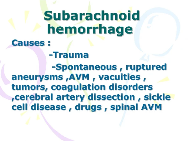
SAH Quick Pathology & Pathophys • Causes • Ruptured aneurysm (75%) • M=F, 5th or 6th decade, acute ∆ BP • Usually congenital berry aneurysms in Circle of Willis • Polycystic Kidney Dz, Coarctation of Aorta, Ehlers-Danlos • HTN, alcohol, cigarettes, cocaine • 2-3% are mycotic aneuryms (s/p infective endocarditis) • Intracranial AVM (10%) • M>F, 2nd to 4th decades • Source of symptoms • Rupture of intracranial artery → ↑ ICP • → distortion of pain-sensitive structures → HA • → decreased cerebral perfusion → LOC • → compression of intracranial structures → 3rd n. palsy, …
History Findings • History • Sentinel/Warning/Thunderclap HA: 20 to 50% get a distinct, unusual, severe HA that precedes the actual HA that causes the pt to seek medical attention; can come days to weeks earlier • Nausea/vomiting • Exertion at time of HA onset • Depressed consciousness • Neck stiffness or pain • Visual changes • Gait disturbance
Physical Findings(Edlow JA, Caplan LR. N Engl J Med. 2000;342(1):29-36) • Nuchal rigidity • Diminished level of consciousness • Papilledema • Retinal and subhyaloid hemorrhage • Third nerve palsy • Sixth nerve palsy • Bilateral weakness in legs or abulias • Nystagmus or ataxia • Aphasia, hemiparesis, or visual neglect
Current Treatment Algorithm(Suarez JI, et al. N Engl J Med. 2006;354(4):387-96) • CT scan without contrast • If positive, perform CT or cerebral angiography* • If negative, perform Lumbar Puncture • If abnormal – CT or cerebral angiography* • If abnormal but equivocal – CT or cerebral angiography* • If normal – Stop * If aneurysm is found, treat promptly. If negative, repeat CT angiogram in 1-3 weeks and image brain, brainstem, and spinal cord.
Sensitivity of 5th generation CT scanners(Boesiger BM, et al. J Emerg Med. 2005 Jul;29(1):23-7) • Retrospective chart review of 177 patients in 2002 that presented with HA and had CT and LP performed to rule out SAH. • Exclusions: trauma within 3 months, age ≤ 17, not having r/o SAH as reason for LP on chart, recent neurosurgery. • Patients were followed up for a minimum of 3 months by chart review and/or phone call to assess for complications after CT and LP were performed • Sensitivity of CT for SAH: 100% (95% CI 61.0-100%) • Specificity of CT for SAH: 99.4% CI 96.8%-99.9%)
Lumbar Puncture(Shah KH, Edlow JA. J Emerg Med. 2002;23(1):67-74) • The gold standard for diagnosis of SAH • ~100% sensitive in detected blood in the CSF • Traumatic tap occurs in ~20% of LPs • Interpretation • “Three tube” test: should see a decrease in traumatic tap vs. steady level of RBCs in true SAH • Xanthochromia: 20% in first 6 hr, 65% between 6 and 12 hr, and 100% after 12 hr • Elevated opening pressure (>20 cm H2O) seen in 60% of cases
Tube 1 Tube 4 Appearance Clear Clear Color None None WBC 0 0 RBC 1 1 CSF Glucose 61 CSF Protein 18 Opening Pressure 16 cm H2O Case: JM • Head CT • Normal • LP results • Phone call follow-up 14 days s/p discharge. • Only one instance of mild HA in past two weeks. • Denies nausea, vomiting, visual disturbance, neck stiffness or any other complaints.
Incorrect diagnoses in misdiagnosed SAH(Edlow JA. Emerg Med Clin N Am. 2003; 21:73-78) • No dx/HA or unknown cause • Primary HA disorder (migraine, cluster, tension) • Meningitis and encephalitis • Systemic infection (flu, gastroenteritis, viral) • Stroke or TIA • Hypertensive crisis • Cardiovascular diagnosis (r/o MI, arrythmia, syncope) • Sinus-related HA • Neck problem (cervical disc dz, arthritis) • Psychiatric dx (alcohol intoxication, malingering) • Trauma-related • Back pain 8 4 Number of episodes required for diagnosis according to Int. HA Society
Misdiagnosis of SAH(Kowalski RG, et al. JAMA. 2004 Feb 18;291(7):866-9) • Inception cohort of 482 SAH patients admitted to Columbia-Presbyterian in NY between 1996 and 2001 • Goal • determine the association between initial missed diagnosis and outcome after SAH • identify factors associated with misdiagnosis • Main outcome measures • Modified Rankin Scale (functional outcome) and Sickness Impact Profile (QOL) at 3 and 12 months (performed by interview in person or via telephone)
Misdiagnosis of SAH(Kowalski RG, et al. JAMA. 2004 Feb 18;291(7):866-9) • Results • Misdiagnosis occurred in 12% (56/482) of patients • Location of initial misdiagnosis • ED (43%) or a physician’s office (32%) • Diagnostic error • No CT performed (73%) • CT or LP results misinterpreted (16%) • CT done, but LP not performed (7%) • Initial misdiagnosis • Migraine/tension HA (36%) • No diagnosis (12%) • Viral syndrome (11%)
Misdiagnosis of SAH(Kowalski RG, et al. JAMA. 2004 Feb 18;291(7):866-9) • Independently associated with misdiagnosis in all patients: • Normal mental status • Small SAH volume • Right-sided aneurysm location • Also associated with misdiagnosis in those presenting with normal mental status: • Education ≤ 12 years • Nonfluency in English • Being unmarried
Case Report #1(Wasserberg J, Barlow P. BMJ. 1997;315(7122):1598-9) • 58M p/w LOC x 1 minute, then had severe HA and hematemesis after awakening • Initial dx: hematemesis • Admitted to hospital, given IM opiates for pain • Initial sx attributed to EtOH withdrawal, pt treated with diazepam • HA not improved after 2 days • SAH was considered, pt booked for elective CT, next appt 2 days later • 1 day before scan, pt became unconscious, had fixed, dilated L pupil • ER CT shows extensive SAH → tx to NICU → died shortly thereafter
Case Report #2(Wasserberg J, Barlow P. BMJ. 1997;315(7122):1598-9) • 17F p/w HA associated with n/v x 1 week • CT to rule out SAH normal → reassured, sent home (CT later reviewed and confirmed normal) • 2 days later she is awakened by sudden HA, she vomits, and collapses • In coma on arrival, reacting to pain only • CT shows SAH, angiogram shows terminal carotid artery aneurysm but patient dies before completion of angiography
Summary • Always consider SAH in a patient who presents with the worst HA of their life • Avoid certain pitfalls: • The patient with known HA history that presents with a new, distinct severe HA • The patient whose clinical picture is complicated by other complaints, intoxication, etc. • Know how to distinguish traumatic tap from SAH to avoid subjecting patients to unnecessary invasive diagnostic testing • Despite advancement in CT scanner technology, today’s data does not support the thought that CT without LP can definitively exclude SAH
References • Boesiger BM, Shiber JR. Subarachnoid hemorrhage diagnosis by computed tomography and lumbar puncture: are fifth generation CT scanners better at identifying subarachnoid hemorrhage? J Emerg Med. 2005 Jul;29(1):23-7. • Coats TJ, Loffhagen R. Diagnosis of subarachnoid haemorrhage following a negative computed tomography for acute headache: a Bayesian analysis. Eur J Emerg Med. 2006 Apr;13(2):80-3. • Edlow JA. Diagnosis of subarachnoid hemorrhage in the emergency department. Emerg Med Clin N Am. 2003 Feb;21(1):73-87. Review. • Edlow JA, Caplan LR. Avoiding pitfalls in the diagnosis of subarachnoid hemorrhage. N Engl J Med. 2000 Jan 6;342(1):29-36. • Edlow JA, Wyer PC. How good is a negative cranial computed tomographic scan result in excluding subarachnoid hemorrhage? Ann Emerg Med. November 2000;36:507-516 • Kowalski BS, et al. Initial misdiagnosis and outcome after subarachnoid hemorrhage. JAMA. 2004 Feb 18;291(7):866-9. • Subarachnoid hemorrhage. Lange Neurology. The McGraw-Hill Companies, 2006. www.accessmedicine.com. • Seehusen DA, Reeves MM, Fomin DA. Cerebrospinal fluid analysis. Am Fam Physician. 2003 Sep 15;68(6):1103-8. • Shah KH, Edlow JA. Distinguishing traumatic lumbar puncture from true subarachnoid hemorrhage. J Emerg Med. 2002 Jul;23(1):67-74. • Suarez JI, Tarr RW, Selman WR. Aneurysmal subarachnoid hemorrhage. N Engl J Med. 2006 Jan 26;354(4):387-96. Review. • Wasserberg J, Barlow P. Lesson of the week. Lumbar puncture still has an important role in diagnosing subarachnoid haemorrhage. BMJ. 1997 Dec 13;315(7122):1598-9. • Wood MJ, Dimeski G, Nowitzke AM. CSF spectrophotometry in the diagnosis and exclusion of spontaneous subarachnoid haemorrhage. J Clin Neurosci. 2005 Feb;12(2):142-6.