Molecular Cell Biology
631 likes | 1.08k Vues
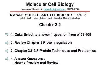
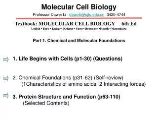
Molecular Cell Biology. Professor Dawei Li daweili@sjtu.edu.cn 3420-4744. Textbook: MOLECULAR CELL BIOLOGY 6th Ed Lodish • Berk • Kaiser • Krieger • Scott • Bretscher •Ploegh • Matsudaira. Part 1. Chemical and Molecular Foundations. Life Begins with Cells (p1-30) (Questions)

Molecular Cell Biology
E N D
Presentation Transcript
Molecular Cell Biology Professor Dawei Li daweili@sjtu.edu.cn 3420-4744 Textbook: MOLECULAR CELL BIOLOGY 6th Ed Lodish • Berk • Kaiser • Krieger • Scott • Bretscher •Ploegh • Matsudaira Part 1. Chemical and Molecular Foundations • Life Begins with Cells (p1-30) (Questions) • Chemical Foundations (p31-62) (Self-review) • (1Characteristics of amino acids, 2 Interacting forces) • 3. Protein Structure and Function (p63-110) • (Selected Contents)
Review • Life Begins with Cells (p1-30) Q&A • Chemical Foundations (p31-62) (Self-review) • 1. Characteristics of amino acids: • 2. Interacting forces:
Chapter 3 Protein Structure and Function (63-110) • 3.1 Hierarchical Structure of Proteins • 3.2 Protein Folding • 3.3 Protein Function • 3.4 Regulating Protein Function I: Protein Degradation • 3.5 Regulating Protein Function II Noncovalent and Covalent Modifications • 3.6 Purifying, Detecting, and Characterizing Proteins • 3.7 Proteomics
Figure 3-1 Overview of protein structure and function.
3.1 Hierarchical Structure of Proteins Figure 3-2 Four levels of protein hierarchy.
The primary Structure of a Protein Is Its LinearArrangement of Amino Acids Figure 3-3 Structure of a polypeptide.
Secondary Structures Are the Core Elements of Protein Architecture The Helix Figure 3-4 The helix, a common secondary structure in protein.
β The Sheet Figure 3-5 The sheet,another common secondary structure in proteins. β
β Turns Figure 3-6 Structure of a turn. β
Overall Folding of a Polypeptide Chain Yields Its Tertiary Structure Figure 3-7 Oil drop model of protein folding.
Different Ways of Depicting the Conformation of Proteins Convey Different Types of Information Figure 3-8 Four ways to visualize protein structure.
Structural Motifs Are Regular Combinations of Secondary and Tertiary Structures Figure 3-9 Motifs of protein secondary structure.
Structural and Functional Domains Are Modules of Tertiary Structure Figure 3-10 Tertiary and quaternary levels of structure.
Structural and Functional Domains Are Modules of Tertiary Structure Figure 3-11 Modular nature of protein domains.
Protein Associate into Multimeric Structures and Macromolecular Assemblies Figure 3-12 A macromolecular machine:the transcription-initiation complex.
Members of Protein Families Have a Common Evolutionary Ancestor Figure 3-13 Evolution of the globin protein family.
3.2 Protein Folding Planar Peptide Bonds Limit the Shapes into which Proteins Can Fold Figure 3-14 Rotation between planar peptide groups in proteins.
Information Directing a Protein's Folding Is Encoded In Its Amino Acid Sequence Figure 3-15 Hypothetical protein-folding pathway.
Molecular Chaperones Figure 3-16 Chaperone-mediated protein folding.
Chaperonins Figure 3-17 Chaperonin-mediated protein folding.
Alternatively Folded Proteins Are Implicated in Diseases Figure 3-18 Alzheimer's disease is characterizes by the formation of insoluble plaques composed of amyloid protein.
3.3 Protein Function Specific Binding of Ligands Underlies the Functions of most Proteins CDR: Complemetarity-Determining Region Figure 3-19 (a) Protein-ligand binding of anti-bodies.
Specific Binding of Ligands Underlies the Functions of most Proteins Figure 3-19 (b) Protein-ligand binding of anti-bodies.
Enzymes Are Highly Efficient and Specific Catalysts Figure 3-20 Effect of an enzyme on the activation energy of a chemical reaction.
An Enzyme's Active Site Binds Substrates and Carries Out Catalysis Figure 3-21 Active site of the enzyme trypsin.
An Enzyme's Active Site Binds Substrates and Carries Out Catalysis Figure 3-22 and for an enzyme- catalyzed reaction.
An Enzyme's Active Site Binds Substrates and Carries Out Catalysis Figure 3-22 (a) and for an enzyme- catalyzed reaction.
An Enzyme's Active Site Binds Substrates and Carries Out Catalysis Figure 3-22 (b) and for an enzyme- catalyzed reaction.
An Enzyme's Active Site Binds Substrates and Carries Out Catalysis Figure 3-23 Schematic model of an enzyme's reaction mechanism.
An Enzyme's Active Site Binds Substrates and Carries Out Catalysis Figure 3-24 Free-energy reaction profiles of uncatalyzed and multistep enzyme-catalyzed reaction.
An Enzyme's Active Site Binds Substrates and Carries Out Catalysis Figure 3-24(a) Free-energy reaction profiles of uncatalyzed and multistep enzyme-catalyzed reaction.
An Enzyme's Active Site Binds Substrates and Carries Out Catalysis Figure 3-24(b) Free-energy reaction profiles of uncatalyzed and multistep enzyme-catalyzed reaction.
Serine Proteases Demonstrate How an Enzyme's Active Site Works Figure 3-25(a) Substrate binding in the active site of typsinlike serine proteases.
Serine Proteases Demonstrate How an Enzyme's Active Site Works Figure 3-25(b) Substrate binding in the active site of typsinlike serine proteases.
Serine Proteases Demonstrate How an Enzyme's Active Site Works Figure 3-26 Mechanism of serine protease- mediated hydrolysis of peptide bonds.
This side-chain of His-57 facilitates catalysis by withdrawing and donationg protons throughout the reaction(inset).
Movements of electrons are indicated by arrows. This attack results in the formation of a transition state called the tetrahedral intermediate
Additional electron movements result in the breaking of the peptide bond,release of one of the reaction products,and formation of the acyl enzyme.
An xygen from a solvent water molecule then attacks the carbonyl carbon of the acyl enzyme.
Additional electron movements result in the breaking of the Ser-195-substrate bond and release of the final reaction product.
Serine Proteases Demonstrate How an Enzyme's Active Site Works Figure 3-27 pH dependence of enzyme activity.
Enzymes in a Common Pathway Are Often Physically Associated with One Another Figure 3-28 Assembly of enzymes into efficient multi -enzyme complexes.
Enzymes in a Common Pathway Are Often Physically Associated with One Another Figure 3-28(a) Assembly of enzymes into efficient multi -enzyme complexes.
Enzymes in a Common Pathway Are Often Physically Associated with One Another Figure 3-28(b) Assembly of enzymes into efficient multienzyme complexes.
Enzymes in a Common Pathway Are Often Physically Associated with One Another Figure 3-28(c) Assembly of enzymes into efficient multienzyme complexes.
3.4 Regulating Protein Function I:Protein Degradation The Proteasome Is a Complex Molecular Machine Used to Degrade Proteins Figure 3-29 Ubiquitin-and- proteasome-mediated prot- eolysis.