Cardiovascular Examination
1.39k likes | 1.79k Vues
Cardiovascular Examination. Deling Zou. Medical ppt. http://hastaneciyiz.blogspot.com. Anatomy. Inspection. 1 Precardial projection and excavation 2 Apical impulse 3 Abnormal pulsations of precardium. Inspection. 1 Precardial projection and excavation 1) Precardial projection

Cardiovascular Examination
E N D
Presentation Transcript
Cardiovascular Examination Deling Zou Medical ppt http://hastaneciyiz.blogspot.com
Inspection 1 Precardial projection and excavation 2 Apical impulse 3 Abnormal pulsations of precardium
Inspection 1 Precardial projection and excavation 1) Precardial projection • congenital heart disease: tetralogy of Fallot • Valvular heart disease-- MS,PS • pericardial effusion (large , childhood)
The second right intercostal space(2nd ICS-RS) • aneurysm of aortic arch • dilatation of ascending aorta 2) flat chest 3) pigeon chest/funnel chest
Inspection 2 Apical impulse *Normal: • position—the fifth left intercostal space 0.5-1.0cm medial to the midclavicular line range—2.0-2.5cm in diameter
*Abnormal1) Location #diaphragm: • “transverse position” upper,outward • obesity ,child, pregnacy; • ascites; tumor of abdominal cavity • “vertical position” (thin, high, emphysema) inferior,inner
#mediastinum: • one side pleural effusion or pneumothorax—to the healthy side • one side atelectesis or pleural adhesion—to the affected
#enlargement of the heart • right ventricular dilatation –left or slightly upper • left ventricular dilatation—left inferior • LV &RV dilatation –left inferior (both side dilatation)
#Posture: • recumbent position—upper • left lateral position—to the left 2-3cm • right lateral position—to the right 1.0-2.5cm • Dextrocardia: 5-ICS—RS
Inspection- apical impulse - abnormal 2)Intensity and extent changes
Inspection -apical impulse - abnormal 3)Inward impulse: • apex excavation in the systole • seen: adhensive pericarditis prominent RV hypertrophy
Inspection • Abnomal pulsations of • percardium 1)left third-forth intercostal space lateral to the sternum(3,4ICS-LS) • seen: RV hypertrophy
2)hypoxiphoid process seen: difference deep inspiration RV hypertrophy ↑ abdominal aorta (aneurysm)↓
3)basal part of the heart • 2 ICS-LS: dilatation of the pulmonary artery or pulmonary hypertensin, occasionally healthy young man • 2 ICS-RS: aneurysm of aortic arch or dilatation of ascending aorta
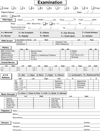
Palpation 1 Apical impulse and pulsation of precardium 2 Thrill 3 Pericardial friction rub
Palpation 1 Apical impulse and pulsation of precardium • Exact position of apex • The beginning of systole of ventricle first sound • Heaving apex impulse: reliable of LV hypertrophy
2 Thrill • One of characteristic signs of organic heart disease. • Mechanism : the flow of blood→narrowed orifice→vortices→ vibration→chest wall • thrill-high frequency murmurs-low frequency • Method:position,phase of cardiac cycle,clinical significance • seen: CHD or valvular stenosis , occasionally insurficiency
3 Pericardil friction rub 1)Precardium-4th ICS-LS 2) both phases of the cardiac cycle 3) systolic period, sitting erect and leaning forward, the end of expiration 4)mechanism: rub of the visceral and parietal layers of pleura 5)seen:acute pericarditis
Percussion • Aim:to determine the size and shape of the heart . • Absolute dullness: contain no gas Relative dullness : real size
1 murneuver of percussion • patient in erect position –the pleximeter is vertical with the intercostal space • patient in the recumbent position –the pleximeter is parallel with the intercostal space
2 order : • left—right ; upwards ; inward • left margin : from 2-3 cm lateral to the apex beat up to the 2nd ICS • right margin : one intercostal space higher than the border of liver dullness up to the 2nd ICS • size: vertical distance from margin to the anterior midline
(2)The upper border –the lower border of the anterior end of the third rib↑ (3)The basal part —the second intercostal space upward left: aortic node and PA (4)Concave part –between the aorta and the left ventricle
Percussion 5 Changes in the area of cardiac dullness and its significance Cardiac factors : 1)LV enlargement: “boot shape” Seen:aortic valvular disease , hypertension heart disease
2)RV enlargement :slightly↑--absolute dullness↑ Prominent↑--relative dullness↑ to the left side prominently Seen:PHD, MS 3)Two ventricle ↑: “generally enlarged heart” seen:DCM , Kashan cardiomyopathy
4)LA and/or pulmonary artery:LA:concave part disappear LA+PA:2,3 ICS-LS outwards “pear shape” Seen: MS--- “mitrial type”
5)pericardial effusion: enlargement of both sides of the border body’s position: • recumbent position:widening of base of the heart • erect position:“triangular shape”
6)dilatation of the aorta /ascending aortic aneurysm: widening if the dull area of first and second intercostal space (with systolic pulsation)
Extacardial factors :1)large pleural effusions and pneumothorax → to the healthy side2)atelectasis /pleural pachynsis →to the affected3)a large amount of ascites or big abdominal tumor: diaphragm elevated→transverse position →left side enlargement
1 Ausclutatoty valve areas 1)ausclutatory mitral area: apical area2)ausclutatory pulmonary area:2 ICS-LS3)ausclutatory aortic area: 2 ICS-RS4)second ausclutatory aortic area: 3rd ICS-LS—Erb area5)tricuspid area :4,5 ICS-LS
2 Order: MV---PV---AV1---AV2---TV 3 Contents:1) rate 2)rhythm 3)heart sound 4)extra heart sound 5)murmurs 6)pericardial friction sound
1)heart rate: • 60~100bpm F>M • child (<3 years) > 100bpm • tachycardia: normal adult >100bpm child(<3 years) >150bpm • bradycardia: HR <60 bpm
Ausclutation heart rate:60-100bmp
2)cardiac rhythm:*sinus arrythmia—affected by breath*premature beat: classification:atrial~ ventricular ~ • junctional ~ • frequently:>6 bpm • occasionally: <6 bpm • bigeminy trigeminy
*atrial fibrillation: absolute irregular rhythm S1 intensity inequality Pulse deficitseen:MS,CHD,hyperthyroidism, PHD,DCM
Ausclutation atrial fibrillation
Ausclutation content cardiac sound • S1: • S2:
4)Abnormal cardiac sound*Intensity: • position of the atrioventricular valve • Ventricular contractility and output • Valvular integrity and activity
S1: Accentuation: • MS • HR↑contractility↑ fever,anemia,hyperthyroidism • complete AVB →cannon sound