EXAMINATION
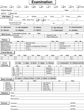
EXAMINATION. Vital Signs. Respiratory Rate- should be counted for a full minute in infants because of variability Heart rate- resting if possible Blood pressure- bilateral upper and lower extremities use correct size cuff! Pulse ox- check/recheck multiple extremities if abnormal reading.

EXAMINATION
E N D
Presentation Transcript
Vital Signs Respiratory Rate- should be counted for a full minute in infants because of variability Heart rate- resting if possible Blood pressure- bilateral upper and lower extremities use correct size cuff! Pulse ox- check/recheck multiple extremities if abnormal reading
Cardiovascular Assessment Is chest symmetrical? Evidence of pectus excavatum, pectus carinatum. Precordial activity quiet or hyperactive? Apex displaced? (usually mid-clavicular line) Palpable thrill over chest? Thrill at suprasternal notch? JVD?
S1 Closure of the atrioventricular valves (mitral and tricuspid) Normally heard as one sound, although occasionally splitting may be heard
S2 Closure of the semilunar valves (pulmonary and aortic) Under normal circumstances, S2 is “split” during or near the end of inspiration (the right ventricle is filled more than the left with inspiration, causing a short delay in closure of the PV) A fixed or widely split S2 could be pathologic, as can a single S2 that does not split.
Murmurs What is a heart murmur?
Innocent murmur ‘• aSymptomatic patient • Soft blowing murmur • Systolic murmur only, not diastolic • left Sternal edge. Also: • Normal heart sounds with no added sounds • No parasternal thrill • No radiation.
Innocent Pulmonary Flow Murmur A relatively soft systolic murmur appreciated at the upper left sternal border Often louder while lying supine and fades/disappears with sitting
Venous Hum Continuous murmur usually heard at the infraclavicular area of the anterior chest, R>L Loudest while sitting (venous return from the jugular veins and subclavian veins entering the SVC ) and disappears supine Can be diminished or muted with gentle compression of jugular vein Commonly associated with vibratory murmur
Diastolic Murmurs Always pathological- there are no “normal” or “innocent” diastolic murmurs What are some diastolic murmurs?
Continuous Murmurs Heard through systole and diastole- most common “pathological” continuous murmur is a Patent Ductus Arteriosus
Cardiovascular Exam (cont’d) Resp- Lungs clear? Equal? Congested? Diminished? Tachypneic? Retractions? Skin- Warm? Pink? Well-perfused? Abdomen- soft? Liver enlarged? Ascites? Situs solititus or inversus? Pulses- Weak? Bounding? Normal? Brachial/radial-femoral delay? Extremities: Capillary Refill Brisk? Clubbing? Edema?
Exam If you want to truly perform a quality cardiovascular examination, auscultate the patient lying, sitting, standing!
The pulse-oximetry monitoring protocol based on results from the right hand (RH) and either foot (F).
Tests of cardiac function Prenatal ultrasound Chest x-ray Electrocardiogram (ECG) Echocardiogram Cardiac catheterization Cardiac MRI CT angio
ECGs in children Heart rate >100 beats/min Rightward QRS axis > +90° T wave inversions in V1-3 (“juvenile T-wave pattern”) Dominant R wave in V1 RSR’ pattern in V1 Marked sinus arrhythmia Short PR interval (< 120ms) and QRS duration (<80ms) Slightly peaked P waves Q waves in the inferior and left precordial leads.
The normal ECG QRS variable with age - Newborn 50-80ms, at 16 years 75-115ms cQTC under 6/12: 490ms, 440ms otherwise Notched t waves; may be normal in V2&3 Transient Wenckebach during sleep
The normal ECG Parameters vary through age Right ventricular dominance owing to high pulmonary pressures, normalise at ~6/12 T waves; usually upright in most leads for first 7/7, then downwards in most leads until adolescence. Upright t waves in childhood may reflect RVH.
Congenital heart disease Foetal PVR>SVR; blood bypasses lungs through ductus arteriosus and foramen ovale DA usually closes 24-36hrs post birth – may be much longer FO closes when left atrium volume increases
Congenital Heart Disease 8-10/ 1,000 liveborn infants will have a congenital cardiac malformation (0.8-1%) Risk of recurrence in families with one parent or one sibling with CHD is 1-4% Some defects are associated with even higher recurrence rates in families. ? Genetics.
What causes CHD? Genetic factors? Environmental factors? Genetic and environmental factors? Possible environmental factors: maternal infection/illness, medication use, substance abuse, chronic diseases such as diabetes, lupus, etc.
Presentation Congenital heart disease presents with: • Antenatal cardiac ultrasound diagnosis • Detection of a heart murmur • Heart failure • Shock. • Cyanosis.
Atrial Septal Defects (ASD) Common defect May be a PFO/small secundum ASD- 20-25% of the population has this May be larger, causing significant L to R shunting Can go undetected for years Exam: wide, fixed splitting of S2, often a systolic murmur r/t increased pulm flow “Easily” fixed (surgery vs. transcatheter)
ECG • Secundum ASD – RBBB RAD • Primum ASD – a ‘superior’ QRS axis (mainly negative in AVF) This occurs because there is a defect of the middle part of the heart where the atrioventricular node is. The displaced node then conducts to the ventricles superiorly, giving the abnormal axis.
Atrioventricular Canal ASD, VSD, and affected mitral & tricuspid valves Associated with Down syndrome Symptoms related to size of holes, degree of valvular involvement, & size of ventricles Often accompanied with pulmonary hypertension
Ventricular Septal Defect Ventricular septum fails to “fill in” completely during embryonic development Various degrees of VSDs from tiny to large May be asymptomatic, mildly symptomatic, or in congestive heart failure May not present clinically until 1-2 months of life Often associated with other lesions Isolated VSD’s typically have favorable surgical outcomes Many small and even mod sized VSD’s can close spontaneously for up to 4 years of age
Ventricular Septal Defect Hole between two ventricles of heart Symptoms related to size & location of VSD and amount of pulmonary blood flow Fix by patching with Goretex
VSD for 30% of all CHD anywhere in the ventricular septum, perimembranous (adjacent to the tricuspid valve) or muscular (completely surrounded by muscle). They can .
Small VSDs These are smaller than the aortic valve in diameter, perhaps up to 3 mm. • Asymptomatic. Physical signs • Loud pansystolic murmur at lower left sternal edge (loud murmur implies smaller defect) • Quiet pulmonary second sound (P2). Investigations CXR • Normal. ECG • Normal.
Large VSDs Symptoms • Heart failure with breathlessness and failure to thrive (faltering growth). • Recurrent chest infections. Physical signs • Tachypnoea, tachycardia and enlarged liver from heart failure • Active precordium • Soft pansystolic murmur or no murmur (implying large defect) • Apical mid-diastolic murmur (from increased flow across the mitral valve after the blood has circulated through the lungs)