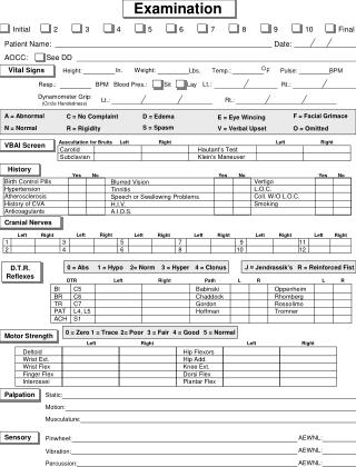
Hematological Examination
Hematological Examination. Jan Živný jzivny@LF1.cuni.cz 22496-5865 / -5934 Department of Pathophysiology. Hematopoietic Cells. Blood / Lymph nodes. Bone marrow. Clinical signs of hematopoietic diseases. Anemi a → hypoxi a → c ardiovas c ul ar signs

Hematological Examination
E N D
Presentation Transcript
Hematological Examination Jan Živný jzivny@LF1.cuni.cz 22496-5865 / -5934 Department of Pathophysiology
Hematopoietic Cells Blood / Lymph nodes Bone marrow
Clinical signs of hematopoietic diseases • Anemia → hypoxia • → cardiovascularsigns • Polycytemia→ hyperviscoseblood • → thrombosis → embolia • → hypoxia • Bleeding → shock →hypoxia • Trombosis → embolia / hypoxia • Frequent infections
Basic: • Complete blood count • Specialized: • Tests for iron metabolism • Erythropoietin measurements • Cytogenetic and known mutation analysis • Immunophenotyping of BM or PB cells • Detection of antibodies to self antigens (e.g. RBC) • Histochemical analysis of cell enzymatic activity • Functional tests (Clonogenic assay) Laboratory Tests
Complete Blood Count (CBC) • Hemoglobin concentration (Hb) • Hematocrit (Hct) • RBC count • RBC parameters • WBC count • WBC differential count • Platelet count • Platelet parameters • Description of blood smear
Hematology Analyzers First automated cell counters came out in the 1950s
How the analyzers work ? Electrical impedance principle (Coulter) cells break an electric circuit as they pass though the aperture between electrodes indicate the presence of a cell (number) and the size of a cell Optical principal – light scatter property of the cell
When to do CBC? • Suspected hematologic, inflamatory, neoplastic, or infection disease • Screening of infants (<1yr.), pregnant women, elderly patients, and patients with nutritional abnormalities • Controversial values during routine patient evaluation
Hemoglobin concentration (Hb) and Hematocrit (Hct) • Depends on age and sex of the patient • Depends on hydratation of the patient (e.g. pregnancy) • F: Hb 121-151 g/L Hct 36-44% • M: Hb 138-170 g/L Hct 41-50% • Less then 70 g/L usually symptomatic tissue hypoxia
ANEMIA • WHO criteria: Hb < 125 g/L in adults • US criteria: • M: Hb < 135 g/L • F: Hb < 125 g/L • Clinical sign
General consequence of anemia hemoglobin loss impaired delivery of oxygen to the tissues tissue hypoxia Symptoms: fatigue, dyspnea, paleness... Signs: tachycardia Anemia
Causes of anemia • Insufficient RBC production: deficient erythropoiesis • Complete loss of erythropoiesis results in Hb decline of about 10% / wk • Excessive RBC loss • Bleeding • Acute: shortly after massive blood loss Hb normal due to vasoconstriction (normochromic - normocytic) • Chronic – leads to depletion of iron which results ininsufficient RBC production • Hemolysis
Red blood cell (RBC) count • F: 3.9 – 5.0 x 10¹² erythrocytes / L • M: 4.5 –5.7 x 10¹² erythrocytes / L
RBC parameters (indices) - 1 Differential diagnosis of anemias MEAN CORPUSCULAR VOLUME = MCV • MCV (fL) = Hct / RBC count • Histological classification of anemias • microcytic anemia ( < 80 fL) • normocytic anemia (80 – 95 fL) • macrocitic anemia (> 95 fL) • Not useful to detect anisocytosis = variation in cell size • Red Cell Distribution Width (RDW 11- 15 %) • Reticulocytosis may increase MCV
RBC parameters (indices) - 2 MEAN CORPUSCULAR HEMOGLOBIN = MCH • MCH (pg/cell) = Hb / RBC count • MCH 32.7 – 33.7 pg / cell • Hypochromia MCH < 27 pg / cell MEAN CORPUSCULAR HEMOGLOBIN CONCENTRATION = MCHC • MCHC (g/L of RBC)= Hb / Hct • MCHC: 267 – 355 g / L
RDW – red cell distribution width Measure the variation of red blood cell (RBC) size (volume) Normal RDW = 11 – 14 % Higher RDW values indicate greater variation in RBC size = anisocytosis Calculated by dividing the standard deviation (SD) of MCV by MCV and then multiplying that result by 100[%]
hypochromic microcytic anemia macrocytic anemia hypersegmented neutrophil
Reticulocyte count • Daily RBC replacement 40,000 – 50,000 /mL • 0.5 – 1.5% of RBC count • Maturate within 1 day in peripheral blood • Criteria of marrow activity • Reticulocytosis • response to blood loss (hemolytic anemias, severe bleeding) • response to therapy of anemia (e.g. B12 or Fe def.) • Reticulocytopenia • deficient erythropoiesis (nutrient , hormonal, etc.) • Reticulocyte index = RI corrects for the severity of anemia RI < 2% indicates hypoproliferative component of anemia RI = Reticulocyte Count x (HCT / normal HCT)
Flow cytometryReticulocyte count Unstained 0.6 – 2.7 % 0 %
Platelet count • 140 – 440 K /mL
WBC count • 4,3 – 10,8 x 10⁹ / L • WBC differential count • Segmented neutrophils: 34-75%; • Band neutrophils < 8%; • Lymphocytes: 12 – 50%; • Monocytes: 3-15%; • Eosinophils < 5%; • Basophils < 3%.
Blood smear • In case of pathologic values in automated analysis of blood count • Morphology of blood elements • Anisocytosis = variation in size • Poikilocytosis = variation in shape (schistocytes=RBC fragments; ovalocytes; spherocytes) • Atypical leucocytes (e.g. blasts)
← sférocyty schistocyty →
Sicle Cell Disease Hemoglobin (Hb) S: záměna valinuza glutamin v pozici 6 Hb beta řetězce
Peripheral blood film chronic myeloid leukemia basophils blast cell
CML leukocytosis with the presence of precursor cells of the myeloid lineage blood film at 400X
CML whole granulocytic lineage, including an eosinophil and a basophil blood film at 1000X magnification
Specialized hematological tests • Tests for iron metabolism • Erythropoietin measurements • Detection of antibodies to self antigens (e.g. RBC) • Cytogenetic and known mutation analysis • Immunophenotyping of BM or PB cells • Histochemical analysis of cell enzymatic activity • Functional tests (Clonogenic assay)
Anemia caused by hemolysis (RBC destruction) reticulocytosis, LDH is increased, unconjugated bilirubin accumulate Extrisic cause (normocytic-normochromic RBC ) • Immunologic abnormalities (AIHA, PNH) • Mechanical injury (trauma, infection) Intrinsic cause • Membrane alterations • congenital (spherocytosis, elliptocytosis) • Aquired (hypophosphatemia) • Metabolic disorders (G6PD deficiency) • Hemoglobinopaties (Sicle cell disease, Thalassemia)
Hemolysis caused by immune mechanisms – Direct Coombs (antiglobulin) test • Detection of antibodies to erythrocyte surface antigens • Antibodies other than to AB antigens • These Abs are responsiblefor hemolysis Antiglobulin serum (anti-hu Ig) is added to washed patientsRBC: agglutination indicates presence of immunoglobulins bound to RBC
Acid hemolysis (Hams’) test • Diagnostic test for paroxysmal nocturnal hemoglobinuria (PNH) • Acidification of blood (HCl) result in hemolysis due to complement activation in PNH patients (Ham) test Urine Blood
Diagnosis of Paroxysmal nocturnal hemoglobinuria (PNH) flow cytometry • Acquired hemolytic anemia due to a hematopoietic stem cell mutation defect • Glycosyl-phosphatidylinositol anchor abnormality caused by the PIG-A gene mutation • Clinical manifestation result from the lack of GPI dependent proteins on the surface of a portion of leukocytes and erythrocytes • Flow cytometry analysis for CD55 and CD59 is used to diagnose PNH
Flow cytometry Technique which allows quantitative and qualitative analysis of cells in suspension. FACS (Fluorescence-activated-cell-sorting) analysis and sorting of cells in suspension based on the differences in light scattering and fluorescence of the cell or cell label.
Měření exprese CD59 průtokovou cytometrií u pacientů s PNH Granulocyty Erytrocyty
Evaluation of hemoglobin metabolism S-bilirubine concentration reflects hem degradation (increase in hemolytic anemia) S-haptoglobin(binds hemoglobin) S-hemopexin(binds hem) (decrease in hemolytic anemia) Hemoglobin electrophoresis identification of abnormal hemoglobins (e.g. hemoglobin S)
Anemia caused by deficient erythropoiesis • Iron deficiency • microcytic-anisocytosis, ↓ reticulocytes • Vitamin B12 or Folate deficiency • macrocytes-anisocytosis • Marrow failure - chronic diseases, aplastic anemia, myelodysplasia, leukemia • normochromatosis-normocytosis • BM hypoplasia
Tests of iron metabolism Serum iron (SI) • F: 600-1400 mg/L, 11-25mmol/L; M: 750-1500 mg/L, 13-27mmol/L • Low in Fe deficiency and chronic disease • High in hemolytic syndromes and iron overload Total iron binding capacity (TIBC) • 2500 – 4500 mg/L , 45-82 mmol/L • High in Fe deficiency • Low in chronic disease Serum ferritin (30-300 ng/mL) • Fe storage glycoprotein • Closely correlates with total body Fe stores • <12 ng/mL Fe deficiency • Elevated in Fe overload, liver injury, tumors (Acute phase protein)
Tests for iron metabolism Serum transferin receptor • Increase in increased erythropoiesis and early Fe deficiency RBC ferritin • storage status over the previous 3 month (Fe deficiency/overload) • unaffected by liver function or acute illness
N ++++ N N ++ Microcytic Hypochromic Anemia (MCV<83; MCHC<31)
0 ++ Microcytic Hypochromic Anemia (MCV<83; MCHC<31)
Serum erythropoietin (EPO) • Enzyme-Linked ImmunoSorbent Assay(ELISA) • Kidney - main source (but some other tissues produce as well) • Expression increased by hypoxia (e.g. caused by anemia) • Expression decreased – kidney disease (ClCr < 45 mL/min), other chronic diseases
1 Antibody coated wells e.g. anti-EPO 2 Antigen binds to antibody e.g. EPO from human plasma/serum A substrate is added and converted by enzyme to colored product 4 3 Second monoclonal antibody binds to immobilized antigen (e.g. EPO) Sandwich ELISA
Bone Marrow (BM) Analysis • Aspiration of bone marrow • usually from posterior iliac crest or sternum 0.5-2.0 mL • Bone marrow biopsy Direct observation of bone marrow activity
Bone Marrow (BM) Analysis • Indication • Unexplained anemia and other cytopenias • Unexplained leukocytosis and thrombocytosis • Suspicion of leukemia and myeloproliferative diseases • Histologic, cytologic, cytogenetic, and molecular biologic analysis
BM biopsy BM failure BM normal
BM cell aspirate: CML BM cells (400X) demonstrates clear dominance of granulopoiesis
Cytogenetic Analysis of BM Chromosomal abnormalities - AML, CML, MDS