Number of released entries
220 likes | 366 Vues
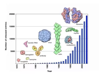
Number of released entries. Year. Growth of Molecular Complexity. Number of Structures Containing that Number of Chains. Number of Chains. Year. HEADER COMPLEX (ACETYLATION/ACTIN-BINDING) 30-MAY-97 1HLU

Number of released entries
E N D
Presentation Transcript
Growth of Molecular Complexity Number of Structures Containing that Number of Chains Number of Chains Year
HEADER COMPLEX (ACETYLATION/ACTIN-BINDING) 30-MAY-97 1HLU TITLE STRUCTURE OF BOVINE BETA-ACTIN-PROFILIN COMPLEX WITH ACTIN TITLE 2 BOUND ATP PHOSPHATES SOLVENT ACCESSIBLE COMPND MOL_ID: 1; COMPND 2 MOLECULE: BETA-ACTIN; COMPND 3 CHAIN: A; COMPND 4 MOL_ID: 2; COMPND 5 MOLECULE: PROFILIN; COMPND 6 CHAIN: P SOURCE MOL_ID: 1; SOURCE 2 ORGANISM_SCIENTIFIC: BOS TAURUS; SOURCE 3 ORGANISM_COMMON: BOVINE; SOURCE 4 ORGAN: THYMUS; SOURCE 5 MOL_ID: 2; SOURCE 6 ORGANISM_SCIENTIFIC: BOS TAURUS; SOURCE 7 ORGANISM_COMMON: BOVINE; SOURCE 8 ORGAN: THYMUS KEYWDS COMPLEX (ACETYLATION/ACTIN-BINDING), ACTIN, PROFILIN, KEYWDS 2 CONFORMATIONAL CHANGES, CYTOSKELETON EXPDTA X-RAY DIFFRACTION AUTHOR J.K.CHIK,U.LINDBERG,C.E.SCHUTT REVDAT 1 15-OCT-97 1HLU 0 JRNL AUTH J.K.CHIK,U.LINDBERG,C.E.SCHUTT JRNL TITL THE STRUCTURE OF AN OPEN STATE OF BETA-ACTIN AT JRNL TITL 2 2.65 A RESOLUTION JRNL REF J.MOL.BIOL. V. 263 607 1996 JRNL REFN ASTM JMOBAK UK ISSN 0022-2836 0070 Challenges in visualization of complexes and the PDB Visualization workshop, October 2003
What is PDB’s role in molecular visualization? Coordinate files of molecules Visualization software = Visualization of molecules +
What is PDB’s responsibility with respect to molecular visualization? providing complete & correctly annotatedcoordinate files in multiple formats (PDB, cif, XML) providing links & explanations for visualization software = visualization for user community – general users, researchers, annotators, students, educators, databases, bio-informaticians +
The PDB format JRNL TITL COMPARISON OF THE THREE-DIMENSIONAL STRUCTURES OF JRNL TITL 2 RECOMBINANT HUMAN H AND HORSE L FERRITINS AT HIGH JRNL TITL 3 RESOLUTION JRNL REF J.MOL.BIOL. V. 268 424 1997 JRNL REFN ASTM JMOBAK UK ISSN 0022-2836 0070 The mmcif format _citation.id primary _citation.title ;Comparison of the three-dimensional structures of recombinant human H and horse L ferritins at high resolution. ; _citation.journal_abbrev J.Mol.Biol. _citation.journal_volume 268 _citation.page_first 424 _citation.page_last 448 _citation.year 1997 _citation.journal_id_ASTM JMOBAK _citation.country UK _citation.journal_id_ISSN 0022-2836 _citation.journal_id_CSD 0070 _citation.book_publisher ? _citation.pdbx_database_id_PubMed 9159481 mmCIF: macromolecular Crystallographic Information File This is an extension of the Crystallographic Information File (CIF) data representation (used for describing small molecule structures) to describe macromolecules.
Complete and correctly annotated coordinate files • Do PDB files conform to uniform standards? • Yes, Remediated mmcif files are available. They can be converted to PDB format using CIFTr (This application is available at http://deposit.pdb.org/software/) • Do PDB files contain coordinates for the complete biological unit? • Yes, both coordinates and pictures of the biological unit of all PDB files are now available.
What is a biological unit? • PDB has coordinates of molecules determined by: • X-ray crystallography • NMR • Electron microscopy • Theoretical modeling Primary coordinate files for crystallographic structure generally contain one asymmetric (unique) unit. • The biological molecule (also called a biological unit) is the macromolecule that has been shown to be or is believed to be functional. This could include one, a part of or multiple asymmetric units.
Concept of the biological unit Biological unit Biological unit could include one, a part of or multiple asymmetric units.
Downloading biological unit images/ coordinate files from the PDB Information for constructing the biological unit is contained in remark 350 of the PDB file PDB ID 1AEW
Visualizing the biological unit Some visualization tools fail to duplicate the secondary structure records for symmetry related molecules in the biological unit Biological unit of 1AEW Viewed in RasMol
3 2 5 Large macromolecular assembly 1: Viruses • For viruses, usually the coordinates of the icosohedralasymmetric unit are deposited to the PDB. Transformation matrices for generating the complete virus are also provided. • Sometimes additional matrices are provided to generate the icosohedral asymmetric unit from the given coordinates Virus particles have high symmetry (5, 3, 2)
Generating the biological unit of a virus Coordinates in crystallographic Symmetry axes Coordinates in icosohedral Symmetry axes Conversion matrix Asymmetric unit NCS applied Asymmetric unit NCS not applied Asymmetric unit 60 matrices Recipe & matrices Crystallographic Symmetry operations Biological unit
Virus: problems and solutions Problems: - matrices for generating the biological unit? - improper nomenclature of the 60 matrices for generating the icosohedral virus particle? - placeholder for the NCS matrices for completing the icosohedral asymmetric unit ? - conversion matrix between crystallographic and icosohedral axes? Possible solutions: - uniform representation of matrices for generating the biological unit? - change in the nomenclature of the 60 matrices for generating the virus? - conversion matrix between crystallographic and icosohedral axes always available? - other?
Large macromolecular assembly 2: Ribosomes • The current PDB format can hold: • a maximum of 99,999 atom records, and • upto 62 different polymer chains. • Since there is no way to represent structures that exceedeither of these restrictions in a single PDB file we have divided such structures into multiple PDB entries. • Although this is not a perfect solution, we have done this to support existing software that rely on current format. • The mmCIF/ XMLformat has no such restrictions.
Ribosomes 1GIX: Small subunit of the ribosome 1GIY: Large subunit of the ribosome
Ribosome: problems and solutions Problems: restrictions in the PDB file format? Size of file? Scaling? Docking? Visualization of nucleic acids? Possible solutions: use of mmCIF/ XML format? better way to represent nucleic acids? other?
What is PDB’s responsibility with respect to molecular visualization? providing complete & correctly annotated coordinate files in multiple formats (PDB, cif, XML) providing links & explanations for visualization software = Visualization for usercommunity – general users, researchers, annotators, students, educators, databases, bio-informaticians +
Annotator needs • Currently we use RasMol and NdbView for quick visualization and checking • For annotation, the visualization software should be capable of: • Quick display (especially for large complexes) • Displaying secondary structure • Selecting atoms or residues for display or rendering • Showing symmetry related molecules • Coloring all or selected residues or chains • Computing distances • Displaying standard and unusual ligands
Educator needs • Visualization programs for educators and students should: • Be free • Be open source • Be capable of running on multiple platforms (without browser dependence) • Be portable • Be easy to install and use • Have user friendly interface Common themes from a survey of ~25 educators from all over the world
Educator requests and visions • Visualization software for educators and students should: • Be more interactive so that students can for example, make mutations in the structure • Have better control on superposition of structures • Have an undo command • Be able to import and export more file formats • Have both a menu driven and command line interface • have different interfaces for research and education and perhaps have a tunable interface • Be a multifunctional suite of programs that can all read the same or related formats
Summary: how do we proceed from here? • How do we ensure that large biological molecules like viruses and ribosomes are uniformly represented in the PDB file? • How do we create a channel of communication between the user community and visualization software developers in order to develop better visualization resources?