Cementum / Periodontium
1.74k likes | 3.99k Vues
Cementum / Periodontium. DEVELOPMENT OF CEMENTUM. Cementum formation is called cementogenesis . Cementoblasts are the cells responsible for cementogenesis . . C ementogenesis. It’s occur right after the enamel was completely formed ;

Cementum / Periodontium
E N D
Presentation Transcript
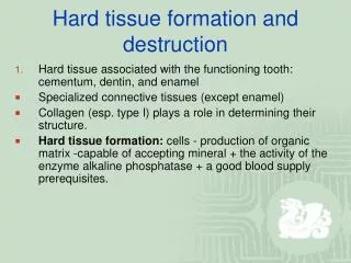
DEVELOPMENT OF CEMENTUM Cementum formation is called cementogenesis. Cementoblasts are the cells responsible for cementogenesis.
Cementogenesis • It’s occur right after the enamel was completely formed; • The outer and inner enamel epithelia together form the epithelial root sheath of Hertwig which is responsible for determining the shape of the root; • The growth of sheath does not occur downward into jaws but that proliferation in cells of sheath causes an upward movement of developing tooth.
These two epithelial layers which are separated by stratum intermedium and stellate reticulum become continous in the area of the future enamel-cementum junction;
Hertwig’s epithelial root sheath • The Hertwig's epithelial root sheath (HERS) is a proliferation of epithelial cells located at the cervical loop of the enamel organ in a developing tooth. • Hertwig's epithelial root sheath initiates the formation of dentin in the root of a tooth by causing the differentiation of odontoblasts from the dental papilla. • The root sheath eventually disintegrates, but residual pieces that do not completely disappear are seen as epithelial cell rests of Malassez (ERM).
Epithelialrootsheath (diaphragm); • Both epithelial layers merged apically into one; • At the bottom form 4-5 cells - guides that do not share; • Cervically begins disintegration of epithelial sheat and begins penetration into mesenchymal cells.
Cementogenesis • Thedifferentiation of odontoblastsfromectomesenchymalcells; • Thefragmentation of Hertwig`sepithelialrootsheath; • Theensuingdifferentiation of cementoblastsfromHertwig`ssheathcellsorfolliclecells.
Cementum is made by cementoblasts • Derived from undifferentiated mesenchyme through intermediate precemntoblast stage in cuboidal cell’s form; • Theyare arranged on outer surface of hyaline layer that covers the dentine.
Cementoblastsare responsible for deposition of organic matrix of cementum which consist proteoglycan and intrinsic collagen fibers. • Also theyareresponsible for subsequent mineralization of organic matrix (cementoid or precementum).
A. HERS did not show any signs of fenestration at this stage; • However,single cells (arrowheads) were positioned between ameloblasts (amel) and the beginning of HERS. • (B). Tooth mineralization was significantly advanced . • The bilayered unit of HERS was still distinguishable at the apex (hers) but had lost its continuity with the ameloblast layer (amel). Instead, bundles of mesenchymal cells (mes, between arrowheads) as well as fibrousstructures (fib) were occupying the developing root surface. Isolated epithelial cells (ep) were localized between mesenchymal tissues and HERS. • Notethe clear separation between ameloblast cell layer (amel, upper row of arrowheads) and mesenchymal cells (mes) covering the developing root surface.
A1 and A2. Note how tubular cells from adjacent to the ameloblast cell layer (arrowheads) were interrupting the continuity of the ameloblast cell layer (amel).B1 and B2. The enamel layer (en) and the ameloblast cell layer (amel) clearly demarked the cervical margin of the developing toothcrown. A membrane (arrowheads) separated the ameloblast cell layer (amel) from the mesenchymal cells (mes) occupying the developing root surface. Therewere only a few epithelial cells (ep) in immediate proximity to the root dentin surface (de)..
Cementogenesis • Cementogenesis occurs when Hertwig`s root sheath disintegrates; • This disintegration of the sheath allows the undifferentiated cells of the dental sac to come into contact with the newly formed surface of root dentin; • This contact of the dental sac cells with the dentin surface induces these cells to become immature cementoblasts.
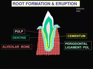
Periodontal ligament and alveolar bone development • As the crown and root develop, the surrounding supporting tissues of the tooth are also developing. The mesenchyme from the dental sac begins to form the periodontal ligament (PDL) adjacent to the newly formed cementum. • This process involves forming collagen fibers that are immediately organized into the fiber bundles of the PDL. The ends of these fibers insert into the outer portion of the cementum and the surrounding alveolar bone to support the tooth. • The mesenchyme of the dental sac also begins to mineralize to form the tooth sockets or alveoli of the alveolar bone surrounding PDL.
(1)Hertwig`ssheat;(2)Epithelialrests of Malassez;(3)Thecells of thedentalfollicle;(4) Cementoblasts;(5)Priodontalligaments;(6) Osteoblasts;(7) Alveolarbone;(8) Odontoblasts.
Mineralization occurs : • When the thin layer of cementoid has formed, mineral salt comes from tissue fluid; • Calcium and phosphate ions will deposited as hydroxyapatite crystal along axes of collagen fibers. • As cementum continues to increase in thinkness, more follicular (extrinsic) fibers become included in cementum, known as Sharpey’s fibers right after periodontal ligament become established.
During the root is developing, those process occurred on apical direction : Proliferation of cells of epithelial root sheath of Hertwigs; Dentine formation; Fragmentation of older part of sheath; Differentiation of new cementoblasts; Formation of cementum.
Cementum • A. Haversian bone ; • B. Bundle bone (A & B make up alveolar bone proper); • C. Reversal line; • D. Osteoid ; • E. Endosteum, composed of osteoblasts ; • F. Cementoid (the organic matrix of cementum) ; • G. Pericementum, composed of cementoblasts.
(A) the position of pulp (pulp), dental follicle (df), mineralized dentin (md),periodontal ligament (lig), Hertwig’s epithelial root sheath (hers) epithelial diaphragm(epd), predentin (pd), dentin (dent), and several blood vessels (bv), • (B) Note the position of epithelial and mesenchymal tissues on the predentin surface at theapical tip of the root. At the apical margin of theroot dentin the periodontal ligament (lig)was in direct contact with the non-mineralizedpredentin (pd). • Hertwig’s root sheath(hers) was separated from the root surface by a periodontal ligament cell layermeasuring at least 10 cell layers in thickness. The nude predentin surface (pd) was notcovered by mineralized dentin or cementum. • At this stage, mesenchymal cells of theligament (lig) had direct access to the root surface. Due to their spatial separation, HERScells had lost their opportunity to deposit cementum on the root dentin prior to theirdeparture from the root surface.
Before tooth erupts :- After tooth erupts : • Extrinsic fiber are incorporated in cementum and lie parallel to the root structure. • The fiber become oblique. • It is known as the precursor of periondontal ligament fibers. Extrinsic fiber
Cementum which is formed first: Does not contain any closed cells, but cementoblast are included in later cementum; These enclosed cementoblast are known as cementocytes and found in lacunae;
Multi-rooted teeth: • Like anterior teeth, multi-rooted premolars and molars originate as a single root on the base of the crown; • This portion on these posterior teeth is called the root trunk; • The root of a posterior tooth divides from the root trunk into the correct number of root branches for its type; • During the formation of the enamel organ on a multi rooted tooth, elongation of its cervical loop occurs in such a way that long, tongue like horizontal epithelial extensions or flaps develop within.
In teeth with more than one root • The initial single primary apical foramen, formed by the epithelial diaphragm of root sheath Hertwig’s, become divided into two or more secondary apical foramina by tongues of epithelial tissue from diaphragm. • These fuse in future furcation area of the roots. The number of secondary apical foramina is determined by the presence of groups of blood vessels which enter the dental papilla.
Two types of cementum form: CellularandAcellular • Acellularcementum forms first. The cementoblasts differentiate from follicular cells, which can only reach the surface of the tooth's root once Hertwig's Epithelial Root Sheath (HERS) has begun to deteriorate; • The cementoblasts secrete fine collagen fibrils along the root surface at right angles before migrating away from the tooth; • As the cementoblasts move, more collagen is deposited to lengthen and thicken the bundles of fibers; • Noncollagenous proteins, such as bone sialoprotein and osteocalcin, are also secreted; • Acellularcementum contains a secreted matrix of proteins and fibers; • As mineralization takes place, the cementoblasts move away from the cementum, and the fibers left along the surface eventually join the forming periodontal ligaments
Cellular cementum Develops after most of the tooth formation is complete and after the tooth occludes (in contact) with a tooth in the opposite arch.This type of cementum forms around the fiber bundles of the periodontal ligaments; The cementoblasts forming cellular cementum become trapped in the cementum they produce; The origin of the formative cementoblasts is believed to be different for cellular cementum and acellularcementum; One of the major current hypotheses is that cells producing cellular cementum migrate from the adjacent area of bone, while cells producing acellularcementum arise from the dental follicle; Nonetheless, it is known that cellular cementum is usually not found in teeth with one root; In premolars and molars, cellular cementum is found only in the part of the root closest to the apex and in interradicular areas between multiple roots.
Cemento enamel junction • In 30% thecementumandenamelmeetas a buttjoint, forming a distinctcementoenameljunctionatthecervicalmargin – edge to edge; • In 60% thecementumoverlapstheenamel; • In 10% have a gap between the cementum and enamel – it lead to sensitivity at this site.
Cementum may be classified in the following ways: • By location: • Radicularcementum: The cementum that is found on the root surface. • Coronal cementum: The cementum that forms on the enamel covering the crown. • By cellularity: • Cellular cementum: Cementum containing cementocytes in lacunae within the cementum matrix. • Acellularcementum: Cementum without any cells in its matrix. • By the presence of collagen fibrils in the matrix: • Fibrillarcementum: Cementum with a matrix that contains well-defined fibrils of type I collagen. • Afibrillarcementum: Cementum that has a matrix devoid of detectable type I collagen fibrils. Instead, the matrix tends to have a fine, granular consistency.
By the origin of the matrix fibers (applies only to fibrillar forms of collagen): • Extrinsic fibercementum: Cementum that contains primarily extrinsic fibers, i.e. Sharpey's fibers that are continuous with the principal fibers of the periodontal ligament. Since the fibers were originally produced by periodontal ligament fibroblasts, they are considered "extrinsic" to the cementum. These fibers are orientated more or less perpendicularly to the cementum surface and play a major role in tooth anchorage. • Intrinsic fiber cementum: Cementum that contains primarily intrinsic fibers, i.e. fibers produced by cementoblasts and that are orientated more or less parallel to the cementum surface. This form of cementum is located predominantly at sites undergoing repair, following surface resorption. It plays no role in tooth anchorage. • Mixed fiber cementum: Cementum that contains a mixture of extrinsic and intrinsic fiber cementum.
CementumVarieties AAC - Acellularafibrillar (prymary) cementum
1. Acellular, afibrillarcementum • This cementum is mostly composed of mineralized matrix, without detectable collagen fibrils or cementocytes. It is produced exclusively by cementoblasts. It is typically found as coronal cementum on human teeth over enamel and dentin in proximity to the cementoenamel junction
2. Acellular, extrinsic fiber cementum This type of cementum has a matrix of well-defined, type I collagen fibrils; The fibrils are part of the, densely packed Sharpey's fibers, that are continuous with the principal fibers of the periodontal ligament; Because of their dense packing, the individual Sharpey's fibers that form the bulk of the matrix may no longer be identifiable as individual fibers within the cementum layer; This cementum, which is acellular, is located in the cervical two-thirds of the root of human teeth; It plays a major role in tooth anchorage.
AcellularExtrinsicFiberCementum (Primarycementum) • It is mainly found oncervical and middle root portions,covering 40% to 70% ofthe root surface; • It serves the exclusivefunction of anchoring the root to periodontal ligament; • Theacellular extrinsic fiber cementum matrix consists of a densefringe of shortcollagenous fibers that are implanted intothe dentinal matrix (glycosaminoglycans) and are oriented about perpendicularly to the root surface; • When theybecome elongated and eventually continuous with theprincipal periodontal ligament fibers they are calledSharpey´s fibers. • Intermingling of collagen fiber bundles with those at the unmineralized dentin. • The final connection between the collagen fiber bundels of acellular cementum and dentin surgace are shown. • They are perpendicular to the dentin and penetrates between the cementoblasts.
3. Cellular, intrinsic fiber cementum • This cementum contains cementocytes in a matrix composed almost exclusively of intrinsic fiber cementum; • It is located at sites of cementum repair.; • It plays no part in tooth anchorage.; • However, it may be covered over by extrinsic or mixed fiber cementum, both of which are able to provide new anchorage; • Once the tooth is in occlusion, a more rapidly formed and less mineralized variety of cementum, cellular intrinsic fiber cementum, is deposited; • Differentiating cementoblast extend cell processes and deposit the collagen fibrils; • As cementum deposition progresses, cementoblasts become entrapped in the extracellular matrix they secrete; • These entraped cells, with reduced secretory activity, are called cementocytes, and the space they occupy is the osteocytic lacuna.
Nourishment of the cells is believed to occur essentially by diffusion, and cementocytes in deeper layers may not be vital.
Collagenfibrilsinthe CIFC Theyaredepositedhaphazardlyduringtherapidphase; Hawever, subsequentlythebulk of fibrilsorganizeasbundlesorientedparallel to therootsurface.
4. Cellular, mixed fiber cementum It is found on the apical third of the root and in furcations (i.e. between roots); In these locations, the rate of cementum formation is usually more rapid than in the cervical region; The mineralized, extrinsic collagen fibers (Sharpey's fibers) run a more irregular course than in acellular, extrinsic fiber cementum; Intrinsic fibers are found interspersed among the extrinsic fibers of the cementum matrix, so that individual Sharpey’s fibers are more readily identifiable than in extrinsic fiber cementum; Cementoblasts are trapped in hollow chambers (or lacunae) where they become cementocytes.