Download
1 / 38
400 likes | 1.49k Vues
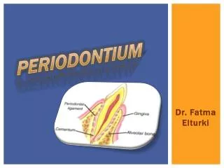
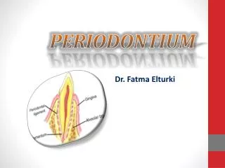
Histology of the periodontium (2) (cont.). Dr. Ahmed Shawkat. The cementum. It is a calcified, avascular, mesenchymal tissue that forms the outer covering of the anatomic root. The two main types of cementum are acellular (primary) and cellular (secondary) cementum .

E N D
Histology of the periodontium (2)(cont.) Dr. Ahmed Shawkat
The cementum • It is a calcified, avascular, mesenchymal tissue that forms the outer covering of the anatomic root. • The two main types of cementum are acellular (primary) and cellular (secondary) cementum. • Collagen fibers in cementum are : • Extrinsic fibers (Sharpey’s fibers) from PDL. • Intrinsic fibers produced by cementoblasts. • Cementum deposition is a continuous process that proceeds at varying rates throughout life. Whereas, the permeability of cementum diminishes with age.
Cementoenamel junction: - Three types of relationships involving the cementum may exist at the CEJ: • In 60 – 65% of cases, the cementum overlaps enamel. • In about 30% an edge-to-edge butt joint exists. • In 5-10% the cementum and enamel fail to meet. In this case, gingival recession can result in accentuated sensitivity because of exposed dentin.
The alveolar bone • Also called the alveolar process, which is defined as parts of the maxilla and the mandible that form and support the sockets of teeth. • Consists of the following: 1- An external plate of cortical bone. 2- The inner socket wall (compact bone) known as alveolar bone proper, seen as the lamina dura in radiographs(continuous radioopaque line) . 3- Cancelloustrabeculae in between. • Both cortical and cancellous alveolar bone are constantly undergoing remodelling(resorption followed by formation) in response to changes in functional forces acting on teeth and age-related changes.
Fenestration and dehiscence: - Isolated areas in which the root is denuded of bone and the root surface is covered only by periosteum and overlying gingiva. When the marginal bone is intact, these areas are termed “fenestrations”. While if the denuded areas extend through the marginal bone; the defect is called “dehiscence”. • They may complicate the outcome of periodontal surgery. • Basal bone: part of maxilla and mandible that supports the alveolar bone proper and that remains after loss of teeth. It tends to be more corticated than the alveolar process.
Clinical criteria of normal gingiva: • Color: the healthy gingiva is usually described as “coral pink” (pale pink), due to the vascular supply, the thickness, and degree of keratinization of the epithelium, and the presence of pigment-containing cells. It is correlated with the cutaneous pigmentation. • The alveolar mucosa is red, smooth, and shiny. Physiologic pigmentation is usually found in black individuals.( due to Melanin, a non-hemoglobin-derived brown pigment).
Form:It depends on the shape and size of interdental area, the gingival margin usually terminate against the tooth in a knife-edge fashion. • The height of the interdental gingiva varies with the location of the proximal contact. • the shape of interdental gingival papillae is correlated with shape of teeth and embrasures: A- Broad interdental papillae. B- Narrow interdental papillae.
Contour: The marginal gingiva envelops the teeth in collar-like fashion and follows a scalloped outline (festooned) on the facial and lingual surfaces. There are interradicular depressions and prominences corresponding to the contours of the roots. • On teeth with pronounced mesiodistal convexity (e.g. maxillary canines) or teeth in labial version, the normal arc-like contour is accentuated, and the gingiva is located farther apically. On teeth in lingual version, the gingiva is horizontal and thickened.
Consistency: The gingiva is firm, resilient, and, with the exception of the movable free gingiva, tightly bound to the underlying bone. • The collagenous nature of the lamina propria determine the firmness of the attached gingiva. Also, the gingival fibers contribute to the firmness of the gingival margin.
Surface texture: The gingiva presents a textured surface similar to an orange peel. The surface of dried gingiva should be matt. The attached gingiva is stippled, while marginal gingiva is not. The central portion of the interdental papillae is usually stippled, but the marginal borders are smooth. • Stippling is a form of adaptive specialization or reinforcement for function. It is a feature of healthy gingiva. It is also related to the presence and degree of epithelial keratinization.
you Thank
Periodontal Instrumentation Dr. Ahmed Shawkat
Classification of Periodontal Instruments • Periodontal Probes • Explorers • Scaling & Root planing Instruments • Cleansing & Polishing Instruments
Scaling & Root Planing Instruments • Curette • Sickel Scaler • File • Chisel • Hoe
Types of Curettes Universal Curette
Types of Curettes Gracy or Area Specific Curettes
Differences between Universal Curettes & Gracy Curettesa • Universal Curette • Gracy Curette
General Principles of Instrumentation • Accessibility • Visibility, Illumination & Retraction • Condition of the Instruments • Mainting a Clean Field • Instrument Stbilization • Instrument Grasping • Finger Rest • Instrument Activation • Adaptation • Angulation • Lateral Pressure
Instrument's Grasping Pen Grasp Modified Pen Grasp Palm & Thumb
Types of Finger Rest Conventional Finger Rest (Intra Oral)
Types of Finger Rest Opposite-Arch Finger Rest (Intra Oral)
Types of Finger Rest Cross-Arch Finger Rest (Intra Oral )
Types of Finger Rest Finger on Finger Rest (Intra Oral)
Types of Finger Rest Palm-up Fulcrum (Extra Oral)
Types of Finger Rest Palm-Down Fulcrum (Extra Oral)
Types of Finger Rest Index Finger Reinforced Rest
Types of Finger Rest Thumb Reinforced Rest