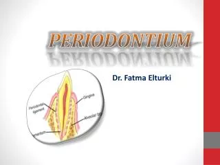
PERIODONTIUM
PERIODONTIUM. Dr. Fatma Elturki. objectives. Periodontium Its definition and composition. Cementum Definition. Physical and chemical characteristics. Development of Cementum. Structure of cementum. Classification of Cementum. Incremental lines of Salter Aging of Cementum.

PERIODONTIUM
E N D
Presentation Transcript
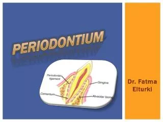
PERIODONTIUM Dr. Fatma Elturki
objectives Periodontium • Its definition and composition. Cementum • Definition. • Physical and chemical characteristics. • Development of Cementum. • Structure of cementum. • Classification of Cementum. • Incremental lines of Salter • Aging of Cementum. • Clinical considerations. • Function of cementum. Periodontal ligament • Definition. • Development of the PDL • Histological structure. • Age changes of PDL. • Functions of the periodontal ligament.
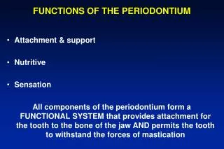
PERIODONTIUM • periodontium is an attachment apparatus of the teeth to the jaws bone. • The periodontium composed of: • Cementum. • Periodontal Ligament. • Alveolar bone. • Gingiva facing the tooth.
Cementum • Cementum is a mineralized dental tissue covering the anatomical part of the root of the human teeth. • It begins at the cervical portion of the tooth at the cementoenamel junction and continues to the apex. • It serves as a medium for the attachment of collagen fibers that bind the tooth to the surrounding structures.
A B Physical characteristics Hardness: Relative softness and the thinness at the cervical portionmeans that cementum is readily removed by the abrasion when gingival recession exposes the root surface to the oral environment. Cementum shares some physical, chemical and structural characteristics with compact bone except that human cementum is avascular and with no nerve supply. Permeability: Cementum has been shown to be permeable to a variety of materials, where it is permeable from the dentin side as well as from the periodontal ligament side. Thickness: the thickness of cementum at the cervical area is about (20-50µm) and it gradually increases in thickness till it reaches its maximum about (150-200u) at the apex and at the bifurcation of the root. Color: cementum is light yellow in color it is somewhat lighter in color than dentin
Chemical Composition In fully formed permanent teeth, cementum contains: • 45% to 50% (inorganic). • 50% to 55% (organic). • The inorganic substances consist mainly of calcium and phosphate in the form of hydroxyapatite. • The organic portion consists mainly of collagen and non-collagenous protein forming the ground substance.
Development of Cementum • Cementum formation occurs along the entire root of the tooth. • Hertwig’s epithelial root sheath (HERS) sends inductive signal to ectomesenchymal pulp cells to secrete predentin by differentiating into odontoblasts. • Once the dentin formation is started, the epithelial root sheath of Hertwig will lose its continuity, undifferentiated mesenchymal cells from adjacent connective tissue of the tooth follicle will differentiate into cementoblasts. • Cementoblasts lay down cementum matrix (cementoid). • Mineralization occurs after some matrix production has taken place.
A-epithelial root sheath B-dental papilla C-odontoblast D-dentine of root E-predentin layer F-cementoblasts G-cementum H-alveolar bone I-inner layer of dental follicle J-outer layer of dental follicle K-intermediate layerof dental follicle
The remnants of the epithelial root sheath of Hertwig migrate toward the dental sac and become the epithelial rests of Malassez found in the periodontal ligament of the fully developed tooth. A-Epithelial rests of Malassez B-cementum
Structure of cementum 1- Cells… • The cells of the cementum are cementoblasts and cementocytes. A-Cementoblasts… • Cementoblasts are mesenchymal cells that form the cementum and are found lining the root surface, interposed between the periodontal fibers. Active cells are round, plump with basophilic cytoplasm and all the organelles associated with protein synthesis. Inactive Cells or resting cells have little cytoplasm and closed nucleus.
B- cementocytes cementocyte is a cell in the lacunae, frequently having long processes radiating from the cell body toward the periodontal surface of the cementum • When acellular cementum is being formed, the cementoblasts resting behind the cementum matrix. When cellular cementum is being formed, the cementoblasts become trapped in lacunae with their own matrix and are then known as cementocytes. Canalicului Lacuna of cementocyte
Structure of cementum 2- The fibrous matrix … The collagen fibers of cementum matrix are of two sorts: • Intrinsic fibersit derived from cementoblasts. Run parallel to the root surface. • Extrinsic fibersit derived from fibroblasts of PDL. These are in the same direction of the PDL principal fibers i.e. perpendicular to the root surface. They are known as Sharpey’s fibers A-cementoblasts B-cementoid C-cementocytes in cellular cementum D-Sharpey’s fiber
Structure of cementum • Under the light microscope two types of cementum can be differentiated: The cementum is usually covered by a zone of cementoid tissue 3-5µm wide in acellular cementum and wider in cellular cementum. This cementoid tissue is lined by cementoblasts. Acellular cementum cellular cementum The difference between the two types is the presence or absence of the cells (cementocytes)
Classification of Cementum Cementum may be classified in the following ways: • By cellularity: Acellular cementum: Cementum without any cells in its matrix. It is clear and structureless it directly overlies the granular layer of Tomes. It covers root dentin starting from the CEJ to the apex, but it is often missing at apical third of the root, where the cementum may be entirely of the cellular type. Cellular cementum: It has the same structure as the acellular cementum but contains cells (cementocyte). It is present in the apical area and overlying acellular cementum. Also common in inter-radicular areas.
Classification of Cementum • By the origin of the matrix fibers: Extrinsic fibercementum: Cementum that contains primarily extrinsic fibers, i.e. Sharpey's fibers that are continuous with the principal fibers of the periodontal ligament. These fibers are play a major role in tooth anchorage. Mixed fiber cementum: Cementum that contains a mixture of extrinsic and intrinsic fiber cementum. Intrinsic fiber cementum: Cementum that contains primarily intrinsic fibers, i.e. fibers produced by cementoblasts. This form of cementum is located predominantly at sites undergoing repair, following surface resorption. It plays no role in tooth anchorage. Acellular cementum Cellular cementum Hyaline layer (of Hopewell Smith) Granular layer of tomes Dentin with tubules
Combined classification Cementum is classified according two factors: • Time of formation (primary or secondary). • Presence or absence of cells within its matrix (acellular & cellular), and origin of collagenous fibers of the matrix (intrinsic fibers and extrinsic fibers). Thus cementum classified into: • Acellular extrinsic fiber cementum (primary cementum) • Cellular intrinsic fiber cementum (secondary cementum). • Mixed fiber cementum where layers of acellular and cellular cementum alternate in apparently random manner. • Acellular afibrillar cementum.
Intermediate cementumHyaline layer of Hopewell-Smith • Sometimes dentin is separated from cementum by a zone known as the intermediate cementum layer. • It is not resemble either dentin or cementum usually it is present in the apical two thirds of the roots of molars and premolars, rarely seen in incisors or deciduous teeth. • It is believed that this layer represents areas where cells of Hertwig's epithelial root sheath become trapped in a rapidly deposited dentin or cementum matrix. • Sometimes the intermediate cementum is found as a continuous layer and sometimes it is found only in isolated areas. A-acellular cementum. B-hyaline layer. C-granular layer of Tomes. D-dentine
Incremental lines of Salter • Both cellular, and acellular cementum are separated by incremental lines into layers, which indicate periodic formation. • Histo-chemical studies indicate that incremental lines highly mineralized areas with less collagen and more ground substance than other portions of cementum. • The incremental lines are roughly parallel to the long axis of the root. • The cellular cementum is formed at a faster rate than the acellular cementum and the incremental lines therefore located further apart than in acellular cementum.
Cementoenamel junction The relation between cementum and enamel at the cervical region of teeth is variable: • In 60% Cementum overlaps enamel. • Cementum just meets enamel in 30%. • 10%Have a small gap between cementum and enamel.
Aging of Cementum • Smooth surface becomes irregular duo to calcification of ligament fiber bundles where they are attached to cementum • Continues deposition of cementum occurs with age in the apical area.[Good: maintains tooth length; bad: obstructs the foramen] • Cementum resorption. Active for a period of time and then stops for cementum deposition creating reversal lines • Resorption of root dentin occurs with aging which is covered by cemental repair
HYPERCEMENTOSIS • It is an abnormal thickening of the cementum. • Hypercementosis may affect one tooth or may be generalized in all teeth, also it is either limited to a small area of the root or through the whole root length. • When the increase in thickness of cementum occurs in a good functioning tooth and it can be considered as a response for the improvement of the functional qualities through increasing the root surface areas and thus permitting more periodontal fibers to be attached to the tooth, this is termed cementum hypertrophy. But if the growth occurs in non functioning teeth or the increase in cementum occurs in embedded teeth this is called cementum hyperplasia.
Cementicles • Calcified ovoid or round nodule found in the PDL. • Single or multiple near the cemental surface. • Free in ligament; attached or embedded in cementum. • common in aging and at sites of trauma. • Origin: Nidus of epithelial cell that are composed of calcium phosphate and collagen to the same amount as cementum.
CLINICAL CONSIDERATIONS • Cementum resorption can occur after trauma or excessive occlusal forces.In most cases of repair, there is a tendency to re-establish the former outline of the root surface by cementum. This is called anatomic repair. • However, if only a thin layer of cementum is deposited on the surface of a deep resorption, the root outline is not reconstructed, & a bay like recess remains. • In such areas the periodontal space is restored to its normal width by formation of a bony projection, so that a proper functional relationship will result. the outline of the alveolar bone in these cases follows that of the root surface. This is called functional repair. Cementum is more resistant to resorption than is bone, & it is for this reason that orthodontic tooth movement is made possible. cementum is similar to bone but has no nerves. Therefore it is non-sensitive to pain. Scaling produces no pain, but if cementum is removed, dentin is exposed causes sensitivity.
Function of cementum • Cementum furnishes a medium for the attachment of collagen fibers of the periodontal ligament to the" alveolar bone. The continuous deposition of cementum is of considerable functional importance, as the most superficial layer of cementum ages, a new layer of cementum must be deposited to keep the attachment apparatus intact. • In the same time deposition of the cementum at the apical part of the root can compensate, to some extent for the loss of tooth substance that may occur from occlusal wear. Cementum serves as a major reparative tissue for root surfaces damage as fracture or resorption can be repaired by deposition of new cementum.
Periodontal ligament • The periodontal ligament is a soft, fibrous specialized connective tissue which is present in the periodontal space, which is situated between the cementum of root of the tooth and the bone forming the socket wall. • It is continuous with the connective tissue of the gingiva and communicates through vascular channels in the bone with marrow spaces. • At the apical foramen it is continuous with the dental pulp. The width of periodontal ligament is not uniform it ranges from 0.15-0.21 mm. The periodontal ligaments of primary teeth are wider than those found in permanent teeth.
Development of the PDL The outer layer of dental follicle (perifollicular mesenchyme) • After separation of HERS, the cells of dental follicle migrate to the external surface of the newly formed root dentin. These migrated follicular cells differentiate into cementoblast that deposit cementum , other cells differentiate into fibroblasts which synthesize the fibers and ground substance of periodontal ligament. The fibers of periodontal ligament become embedded in the developed cementum and alveolar bone. As the tooth erupts the fibers of the periodontal ligament are oriented in the characteristic fashion. The inner layer of dental follicle dental follicle Developing tooth
Periodontal ligament (PDL) Only after the teeth come into occlusion do the fiber bundles of PDL become well organized in terms of thickness and orientation At first the developing fiber bundles of the PDL are thin and not well oriented. They are all directed obliquely
Histological structure • Similar to all connective tissue the PDL consists of: Extracellular substances Cells Fibers Synthetic Ground Substance Resorptive BV Progenitor lymphatic Defensive Nerve
I- The cells Synthetic cells fibroblasts, osteoblasts & cementoblasts. Resorptive cells Fibroblasts, osteoclasts & cementoclasts. epithelial cells remnants of the epithelial root sheath of Hertwig Progenitor cells undifferentiated mesenchymal cells Defensive cells macrophage, lymphocytes &mast cells
II- The fibers *The fibers of the periodontal ligament are mainly collagen. They are divided into: A) The principal fibers. B) The oxytalan fibers. *Elastic fibers are restricted almost entirely to the walls of blood vessels.
A- The principal fibers 1- principle fiber bundle of the PDL: • The alveolar crest group. • The horizontal group. • The oblique group. • The apical group. • The interradicular group.
A- The principal fibers • At the end, all the principle collagen fiber bundles of the PDL are embedded in cementum or bone. The embedded portion is referred to as Sharpey’s fiber.
A- The principal fibers 2- Gingival ligament:- They are found in the lamina propria of the gingiva: • Dentogingival group. • Alveologingival group. • Circular group. • Dentoperiosteal group. • Transseptal fiber system.
These are immature elastic (pre-elastic) fibers. They need special stains to be demonstrated. They tend to run in an axial direction, one end being embedded in bone or cementum and the other in the wall of blood vessels. At the apical region they form a complex network. The function of the oxytalan fibers has been suggested that they supporte the blood vessels of the periodontal ligament during mastication i.e., it prevents the sudden closure of the blood vessels under masticatory forces. B- Oxytalan fibers
Interstitial tissue • The ground substance fills the spaces between cells, fibers, • They are areas containing some of the blood vessels, lymphatics and nerves and surrounded by loose connective tissue. Ground Substance
Blood supply Main blood supply is from superior and inferior alveolar arteries. The blood vessels are derived from the following: 1. BRANCHES FROM APICAL VESSELS Vesselssupplying the pulp. 2. BRANCHES FROM INTRA-ALVEOLAR VESSELS:- Vessels run horizontally and penetrate the alveolar bone to enter into the periodontal ligament. 3. BRANCHES FROM GINGIVAL VESSELS:- The arterioles and capillaries ramify and form a rich network. Rich vascular plexus is found at the apex and in cervical part of ligament.
(A) Subepithelial capillary network of the gingiva, (B) Capillary network of the periodontal ligament, (C) Supraperiosteal arterial, and (D) Arterials penetrating the interdental alveolar bone. Lymphatics: A network of lymphatic vessels follows the path of blood vessels. Usually the flow is from the ligament toward and into the adjacent alveolar bone.
NERVE SUPPLY • Nerves found in ligament pass through foramina in alveolar bone. • The nerves are the branches of second and third division of fifth cranial nerve (trigeminal nerve) and follow same path as blood vessels. • These nerve fibers provide sense of touch, pressure, pain and proprioception during mastication.
The Age Changes of periodontal ligament *The periodontal ligament through aging shows: vascularity cells thickness *They may contain cementicles.
Functions of the periodontal ligament The periodontal ligament has the following functions: • Supportive: Periodontal ligament forms a functional system, which provides an attachment for the tooth to the bone of the jaw, while at the same time permitting the teeth to withstand the considerable forces of mastication. • Sensory: The periodontal ligament having the mechanoreceptor contributes to the sensation of touch and pressure on the teeth. So the proprioceptive reflex protects the tooth in case of sudden overload. It causes inhibition of the activity of the masticatory muscles. Opening the mouth at once relieves the pressure.
Functions of the periodontal ligament • Nutritive: The blood vessels in the periodontal ligament provide nutrient supply required by the cells of the ligament and to the cementocytes and the most superficial osteocytes. • Formative: The cells' present in the periodontal ligament as the fibroblasts are responsible for the formation of new periodontal ligament fibers and dissolution of the old fibers as the continued adaptive readjustment of the suspensory ligament demands. Another formative function is fulfilled by the cementoblasts and osteoblasts which are essential in building up cementum and bone. • Protective: The protective function of the periodontal ligament is achieved by: a- The principal fibers. b- The blood vessels, c- The nerves.
Thank you summary