
SlideServe
Menu
Préférences
© SlideServe · v2026

Understanding Estrous Cycle Stages in Rats Using Vaginal Smear Techniques

cher avatar
6 diapositives · 200 Views · Oct 21, 2014

This guide outlines the procedure for assessing the estrous cycle in rats through vaginal smears. It details the characteristics of each estrous phase, including Diestrous, Proestrous, Estrous, and Metestrous, highlighting the presence of epithelial cells and white blood cells. A step-by-step protocol for obtaining samples, staining them, and analyzing the results under a microscope is provided to help researchers accurately determine the estrous phase and maintain proper records for future reference.
Understanding Estrous Cycle Stages in Rats Using Vaginal Smear Techniques - slide 1
01 / 6Understanding Estrous Cycle Stages in Rats Using Vaginal Smear Techniques
1 / 17

Télécharger la présentation

Understanding Estrous Cycle Stages in Rats Using Vaginal Smear Techniques
Une image / un lien ci-dessous est fourni (tel quel) pour télécharger la présentation

Politique de téléchargement : le contenu du site vous est fourni TEL QUEL pour votre information et votre usage personnel ; il ne peut être vendu, concédé sous licence ni partagé sur d’autres sites sans l’accord de son auteur.Le contenu vous est fourni TEL QUEL, pour votre information et votre usage personnel uniquement. Téléchargez la présentation en cliquant sur ce lien. Lors du téléchargement, si vous ne parvenez pas à télécharger une présentation, l’éditeur a peut-être supprimé le fichier de son serveur.Pendant le téléchargement, si vous ne parvenez pas à obtenir une présentation, le fichier a peut-être été supprimé par l’éditeur.

F I N

Plus de contenu associé

Transcription de la présentation
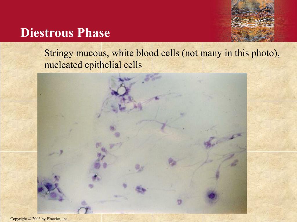
  1. Diestrous Phase Stringy mucous, white blood cells (not many in this photo), nucleated epithelial cells

  2. Proestrous Mostly nucleated epithelial cells

  3. Early Estrous Relatively even distribution of nucleated epithelial cells and cornified epithelial cells

  4. Estrous Mostly cornified epithelial cells

  5. Metestrous Many WBC, some nucleated and cornified epithelial cells

  6. Vaginal smear procedure: • Use clean pipet tip and inject about 25 microliters of 0.9% saline into the vagina of the rat. • With same tip, obtain a sample of the vaginal lavage and transfer to a clean slide (label with initials and the date). • Let dry, then stain for approximately 30-45 seconds in Wright stain (place a drop or two of stain on your slide). • Rinse slide briefly in water, then observe smear under microscope. Record description of smear and determine estrous phase. Retain slide for future reference if needed (store in labeled box in animal room).