Download
1 / 52
560 likes | 1.79k Vues
Mitochondrion and Chloroplast Ultrastructure. Discovery of Mitochondria. Kollicker (1850) teased mitochondria from striated muscle Altmann (1890) used special stains to identify these granules and named them bioblasts

E N D
Discovery of Mitochondria • Kollicker (1850) teased mitochondria from striated muscle • Altmann (1890) used special stains to identify these granules and named them bioblasts • Warburg (1910) used low-speed centrifugation to isolate mitochondria and showed it contained enzymes for catalysing oxidative reactions • 1950 Lehninger, Green and others clearly showed that fatty acid oxidation and oxidative phosphorylation were properties of mitochondria
Structure of Mitochondria • The number and distribution of mitochondria in a cell are closely related to the activity of the cell. Eg cells undergoing movement may display increased numbers of mitochondria • Most are ovoid bodies with a diameter of between 0.5 – 1.0 micrometers and a length of up to 7 micrometers
Structure of Mitochondria • Outer membrane- contains special pores which make it freely permeable to most small molecules and ions • Matrix-gel-like solution which contains enzymes, coenzymes responsible for fatty acid oxidation etc. • Inner membrane-is specialized highly convoluted membrane which is impermebale to most small ions like H+, ATP, ADP • Specialized transport systems are necessary to move ions across the membrane
ATP SYNTHETASE COMPLEXES • These are protein complexes referred to as inner membrane particles and are attached to the inner surface of the inner mitochondrial membrane They appear as spheres that protrude into the mitochondrial matrix
Matrix of the Mitochondria • Gel-like solution contains enzymes responsible for the oxidation of pyruvate, amino acids, fatty acids and those of the TCA cycle • The synthesis of urea and heme occur partially in the matrix • Contains coenzymes NAD+ and FAD (the oxidized forms of the 2 coenzymes that are required as hydrogen acceptors • Contains ADP and Pi which combine to produce ATP
Electron Transport Chain • Located in the inner mitochondrial membrane • Energy-rich molecules are metabolized by a series of oxidation reactions to yield CO2 andH2O • The metabolic intermediates donate electrons to specialized coenzymes NAD+ and FAD to form energy rich NADH and FADH2. These in turn donate a pair of their electrons to a special set of electron carriers collectively called the ELECTRON TRANSPORT CHAIN • As electrons move down the chain they lose free energy, part of this energy is used in the production of ATP. This process is called OXIDATIVE PHOSPHORYLATION
Organization of the Chain • The inner mitochondrial membrane can be disrupted into five separate enzyme complexes Complex IComplex IV each contain the electron transport chain Complex V catalyzes ATP synthesis • Each complex Accepts OR Donates electrons to the next carrier in the chain • The end result is a combination of protons with oxygen to form water • This requirement for oxygen makes the electron transport chain O2 utilization the greatest in the body
Reactions of the Electron Transport Chain • All members of the chain excepting Coenzyme Q (ubiquinone) are protein • May be enzymes eg dehydrogenases • May contain iron as a part of an iron-sulphur center • May be coordinated with an porphyrin ring like the cytochromes • May contain copper eg cytochromes a+a3
Reactions of the Electron Transport Chain • Formation of NADH • NADH dehydrogenase enzyme complex • Coenzyme Q • Cytochromes B and C • Cytochrome a+a3
1. Formation of NADH • NAD+ is reduced to NADH by dehydrogenases which remove 2 Hydrogen atoms • Both electrons but one proton (H- hydride ion)are transferred to the NAD+ to form NADH
2. NADH Dehydrogenase • The free proton plus the hydride ion carried on NADH are transferred to NADH dehydrogenase (NADH-Q reductase) • Enzyme complex embedded on the inner membrane • FMN, a coenzyme accepts the 2 hydrogen atoms and forms FMNH2
Coenzyme Q • Coenzyme Q can accept both hydrogen atoms from FMNH2 and FADH2. FADH2 is produced by succinate dehydrogenase and acyl CoA dehydrogenase
Cytochromes b,c and a+a3 • Cytochromes contain a heme group and a porphyrin ring • Electrons are passed down the chain from coenzyme Q to cyochromes b,c and a+a3 • Cytochrome a+a3 is the only electron carrier in which the heme iron has a free ligand and can react directly with molecular oxygen • Transported electrons bind with oxygen to produce water
Inhibitors of electron transfer • Rotenone and amytal block electron transfer in the NADH-Q reductase • Antimycin A blocks from cytochrome b reductase • CN-, N3- and CO block cytochrome oxidase by binding with the heme at a3
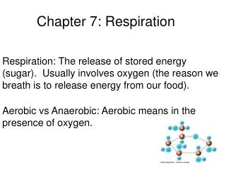
ATP Formation • Produced in the cytosol during glycolysis in which sugars are catabolized • May be produced within chloroplasts by the utilisation of light energy • May be produced within the mitochondria present in all plant and animal cells by the oxidation of elementary substrates
Oxidative Phosphorylation • The process by which ATP is formed as a result of the transfer of electrons from NADH or FADH2 to oxygen by a series of electron carriers • THIS IS THE MAJOR SOURCE OF ATP • Generates 26 of the 30 molecules of ATP formed when glucose is completely oxidized to CO2 and H2O
Electron motive force is converted to proton-motive force and then into phosphoryl potential • Oxidation and phosphorylation are coupled
NADH +1/2 O2 +H+ -H2O +NAD+ • ΔG°’ = -52.6 kcal/mol • ADP+Pi+H+-ATP+ H2O • ΔG°’ = +7.3 kcal/mol
The Proton Pump • Electron transport is coupled with the transport of protons across the inner mitochondrial membrane from the matrix to the inner membrane space • This creates an electron gradient with more +ve charges outside the membrane • A pH gradient is also created, the outside of the membrane is at a lower potential than the inside • The energy generated is sufficient to drive ATP synthesis
Chemiosmotic Hypothesis • After the protons have been transferred to the cytosolic side of the inner mitochondrial membrane they reenter the mitochondrial matrix by passing through a channel in the ATP synthetase molecule • Thus forming ATP from ADP and Pi • ATPase spheres can be seen on the inner mitochondrial membrane and are referred to as F1 • F1 unit catalyzes the synthesis of ATP
The other major unit of ATP synthase is Fo, • A hydrophobic segment which spans the inner membrane • Is the protein channel of the complex • The stalk between F1 and F0 is sensitive to oligomycin
ATP Synthesis • The enzyme complex ATP synthetase (ATPase) synthesizes ATP utilising the energy generated by the proton motive force of the electron transport chain • Enzyme bound ATP forms without the presence of the proton motive force • The proton gradient releases ATP from the synthase complex • Catalyzes the formation of ATP from ADP and orthophosphate (Pi)
Electrons from cytosolic NADH enter mitochondria by shuttle • Electrons from NADH rather than NADH itself are carried across the mitochondrial membrane • Glycerol phosphate shuttle • Malate aspartate shuttle
The entry of ADP into Mitochondria is coupled to the exit of ATP by the ATP-ADP translocase • ADP only enters the mitochondrial matrix if ATP exits and vice versa • Mitochondrial transporters for metabolites have a common tripartite motif
THE Rate of oxidative phosphorylation is dependent on the need for ATP • This is determined by the level of ADP • The rate of O2 consumption increases when ADP is added then returns to its original value when ATP has been synthesized • Electrons do not flow from fuel molecules unless ATP needs to be synthesized
ATP yield • Glycolysis 2 • Citric acid cycle 2 • Oxidative phosphorylation 26 • TOTAL 30
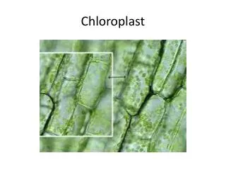
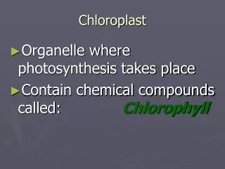
ALL free energy for biological systems arises from solar energy by a process known as PHOTOSYNTHESIS • Photosynthesis takes place in chloroplasts • Chloroplast are 5 micrometers long • Like mitochondria they contain an outer and an inner membrane and an intermembrane space
The inner membrane surrounds a stroma, containing soluble enzymes • Membranous structures called thylakoids (flattened discs) • A pile of thylakoids is called a granum • Thylakoid membranes contain energy transducing machinery: the light harvesting proteins, reaction centers electron transport chains and ATP synthase
The outer membrane is permeable to small ions and molecules • The stroma contains soluble enzymes that utilise NADPH and ATP synthesized by the thylakoids to produce sugar
Chlorophyll • The principal photoreceptor • Is a magnesium porphyrin whereas heme is an iron porphyrin • Have very strong absorption bands because they contain networks of alternating single and double bonds called polyenes
REACTION CENTER • Photosynthesis can be separated into light and dark reactions • The light reactions generate NADPH and ATP • The dark reaction use NADPH and ATP to reduce CO2
Transmembrane proton gradient • Electon flow from PII to PI as well as within the system generates a transmembrane proton gradient that drives ATP formation • Photosystem II transfers electrons from water to plastoquinone • Plastiquinone resembles ubiquinone (CoQ)
D1 and D2 • Phtosystem II is formed by D1 and D2 a pair of subunits that span the thylakoid membrane • D1 and D2 contain the reaction center and the electron transfer chain
Electron Transfer PI • Electronic Excitation of P680 • Binds to pheophytin • Binds to plastoquinone at the Qa site • Reduces plastoquinone to quinone • Binds to Qb site • Electron flows from H2O to QH2 • Formation of a proton gradient as the electron flows through cytochrome bf complex catalyses the transfer of electrons plastoquinol to (QH2) to plastocyanin and concomitantly pumps protons across the thylakoid membrane
Electron transfer PI • An electron is transferred from P700 (reaction site of PI) • P700 gets excited • The electron is transferred from P700 to Ao ( a monomeric acceptor chlorophyll) • AoAo- and P700+ (most potent reductant in biological systems • P700+ captures an electron from plastocyanin P700 • Ao- is transferred to A1 a quinone and hen to Fx an iron-sulphur cluster • FxFerredoxin • Net rxn PI is PC (Cu+)+ ferredoxin (oxidized)-->PC (Cu++) +ferredoxin (reduced)
NADPH Formation • High potential electrons of ferredoxin are transferred to NADP+ to form NADPH • This rxn is catalyzed by a flavoprotein ferredoxin-NADP+ reductase • The net rxn of PII is • 2H2O + 2 NADP+ O2 +2NADPH + 2H+ • Light causes electrons to flow from H2O to NADPH and leads to the generation of a proton-motive force
ATP synthesis • Driven by a proton-motive force in both photphosphorylation and oxidative phosphorylation • ATP synthase is very similar to mitochondria the complex is called CFo-CF1 • Recurring motifs and mechanisms in photosynthetic reaction centers
The Reaction center • P680 is a reaction center