Download

1 / 90
920 likes | 1.27k Vues
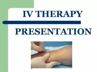
Basic IV Therapy and review of Phlebotomy. Megan Kennedy Cochise County Jail Medical. Agenda for the day……. Discuss purpose and uses of class Anatomy & physiology Identifying the purposes of IV infusions IV solutions Setting up an IV IV catheters Selecting the IV site

E N D
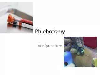
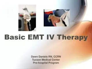
Basic IV Therapy and review of Phlebotomy Megan Kennedy Cochise County Jail Medical
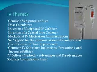
Agenda for the day……. • Discuss purpose and uses of class • Anatomy & physiology • Identifying the purposes of IV infusions • IV solutions • Setting up an IV • IV catheters • Selecting the IV site • Starting the IV • Complications • Trouble-shooting • Removing the IV line • Drip calculations • Phlebotomy side of things
This course is designed to develop the following course competencies: • Identify the need for fluid resuscitation in pediatric and adult victims • Identify and describe the vascular anatomy and venous access for the pediatric or adult victims • Identify and differentiate isotonic, hypotonic, and hypertonic solutions; • Select fluids, set up and manage equipment • Identify and demonstrate aseptic and safety techniques • Identify and describe indications and contraindications for intravenous site selection • Perform all peripheral intravenous cannulation techniques, monitor infusion • Demonstrate 100% accuracy in intravenous techniques in selected scenarios • Demonstrate 85% proficiency on a written examination. • Discuss how fluid resuscitation pertains to phlebotomy. • Continue or begin your education in intravenous access.
Introduction • Knowing the anatomy will aid you in performing your skills, even when you cannot see the veins. • After this block of instruction you should be able to differentiate between veins and arteries, and show where these items can be found.
Skin • Covers the entire body and acts as a protective layer between the body and the environment. • The main functions of the skin are: • Protection from harmful influences • Control of body temperature • Conveyance of sensory impressions • Some areas of the bodies skin are highly sensitive and the insertion of a needle in one area may cause a great deal of pain, while another area may be practically painless.
Besides epithelial cells and connective tissue cells, the skin also contains delicately entwined nerves and blood vessels.
Blood Vessels • With the exception of capillaries, the walls of the blood vessels consist of three layers, though the thickness or construction of the individual layers can vary according to the vessel type.
Blood Vessels • The outer layer consists of connective tissue and facilitates the fitting of the vessel into its environment. • The middle layer is composed of smooth muscle containing elastic fibers. • The inner layer consists of thin connective tissue. It is covered by a layerof single-layered endothelial cells.
Arteries • The arteries are blood vessels which transports blood away from the heart. • They are different in construction from the veins in that they have an additional layer of an elastic membrane situated between the inner and middle wall layers.
Depending on the task and the location of the artery, its middle layer may be dominated by smooth muscle or elastic fibers. Arteries
Arteries • When the heart pumps blood into the arteries during the expulsion phase (systole), their high proportion of elastic fibers permits them to distend. • During the relaxation phase (diastole) of the heart, they contract again, transporting blood on further. • Arteries with muscle predominating are able to widen (vasodilation) or narrow (vasoconstriction) their diameter through contraction, thus enabling the amount of blood contained within them to increase or decrease with the demands of the body. • Arterioles are the smallest arteries.
The veins are the blood vessels which transport blood towards the heart. The wall layers of the veins are thinner than those of the arteries, yet contain more connective tissue. The muscle layer is less marked. The diameter of veins are larger than that of arteries. Veins
Veins • As a result of the thin layer of muscle the veins are not able to move blood themselves. They are aided by the surrounding musculature around them. • In order to prevent the blood from flowing back, some of the veins, especially those within the extremities, are equipped with venous valves. • When the blood is flowing towards the heart, the venous valves lie flat against the venous wall. If the blood congests or starts to flow back, the valves close.
Capillaries • Capillaries are the smallest blood vessels in the body • They are connected to the arterioles and into the venules, thus representing the link between arteries and veins.
Capillaries • In contrast to arteries and veins, capillaries have neither a middle or outer wall layer. They only have an inner layer, constructed of connective tissue and endothelial cells. • The diameter of capillaries is very small because of they are so small they circulate in single file.This fact, and the thinness of their wall layer promote their ability to exchange material and Water with their environment. • The oxygen and nutrients contained within the blood are pressed out of them as a result of the blood pressure and passed off to the intercellular cavities. • Carbon dioxide and metabolic products are absorbed by the blood in the exchange
Blood • 4-5x thicker than water • Liquid connective tissue • Adults = 7% of patients weight • 4-6 Liters of blood • Children = 9% of patients weight
Physical Characteristics of Blood • Color range • Oxygen-rich blood is scarlet red • Oxygen-poor blood is dull red • pH must remain between 7.35–7.45 • Blood temperature is slightly higher than body temperature (100.4)
Functions of Blood • Transportation – oxygen, nutrients, hormones, heat, electrolytes. • Carries away from the body tissues - Waste matter - CO2 • Protection – Vital role in our immune system; clotting mechanisms that prevent blood loss • Regulation – pH, body temperature, water content
Components of Blood • PLASMA – is the yellowish fluid of the blood and consists primarily of water (92%) and plasma proteins (7%) • Proteins – albumin and fibrinogen • FORMED ELEMENTS – solid component of the blood consisting of red blood cells, white blood cells, and platelets • BLOOD = 55% plasma
Blood Plasma • Composed of approximately 92 percent water • Contains: • Nutrients, salt solution • Respiratory gases • Hormones • Proteins, Waste products
Plasma Proteins • Albumin – regulates osmotic pressure • Clotting proteins – help to stem blood loss when a blood vessel is injured • Antibodies – help protect the body from antigens
Formed Elements“Types of Cells” • Erythrocytes = Red Blood Cells • Leukocytes = White Blood Cells • Thrombocytes = Platelets
Erythrocytes “Red Blood Cells” • The main function is to carry oxygen • Anatomy of circulating erythrocytes • Cells without a nucleus • Produced continuously in the bone marrow from stem cells at a rate of 2-3 million cells per second. ─ Hemoglobin 95% of a red cell • Approximately 120 days life span • Outnumber white blood cells 1000:1
Leukocytes“White Blood Cells” • These are complete cells, with a nucleus • Crucial in the body’s defense against disease. Ingest pathogens & destroy • Produce antibodies • Can respond to chemicals released by damaged tissues
Thrombocytes“Platelets” • Cell fragments without nuclei that release blood clotting chemicals • Life span of 5-9 days • Needed for the clotting process • Platelets and clotting proteins work together.
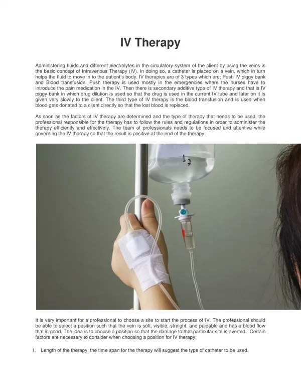
Purpose of starting IV’s • To deliver fluids • To deliver medications
Cellular Physiology • Body Fluid • 1. Total body water = 60% of body weight • 2. Electrolytes • Sodium • Potassium • Magnesium • Calcium • Bicarbonate • Chloride
3. Protein • Albumin • Plasma • Other • FLUID LOSS: • Blood loss • Plasma loss • Nausea/vomiting/diarrhea • Sweating
Isotonic – IV fluids that approximate the osmolaity of blood plasma. I.e.: 0.9% Normal Saline (note the biconcave shape of the cells as they circulate in blood.) Hypotonic – IV solutions that have a lower osmolarity than blood plasma thus drawing fluids into the cell. I.e.: D5W. (note the cells are visibly swollen and have lost their biconcave shape, and at 100 mOs, most have swollen so much that they have ruptured, leaving what are called red blood cell ghosts. In a hypotonic solution, water rushes into cells.) Hypertonic – IV fluids that have a higher osmolality than normal blood plasma thus drawing fluids out of the cells and they get irritated when infused.I.e.: D50 (note water has flowed out of the cells, causing them to collapse and assume the spiky appearance you see.)
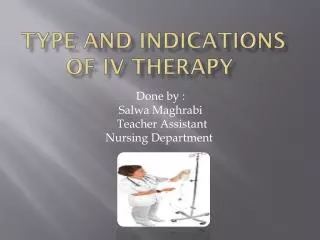
Types of IV Solutions • 0.9% Sodium Chloride • Lactated Ringers • D5W
0.9% Sodium Chloride • Also called normal saline • Isotonic solution of sodium chloride in water • 9 grams of sodium chloride per liter • Indications • Restore loss of water and sodium chloride • Fractures • Trauma • Dehydration • Hypoglycemia • Non-traumatic hypoperfusion • Contraindications • Use with caution in CHF and pulmonary edema
Lactated Ringers • Source of water, electrolytes, and calories • Indications • To replenish fluid and calories, and restore loss of electrolytes • Trauma • Burns • OB • Non-traumatic hypotension • dehydration • Contraindications • Use with caution in CHF, pulmonary edema, and liver disease
D5W • Hypotonic solution of dextrose in water (50 grams of dextrose per liter) • Indications • Directed by MD • Contraindications • Head injury • Children
Combinations of Normal Saline, Lactated Ringers, and D5W are often common. All fluids come in 250cc, 500cc, and 1000cc bags.
Assemble and prepare the necessary equipment You will need: The correct IV solution The correct administration set An IV catheter An IV start pack Tourniquet Alcohol prep Opsite or equivalent Tape
Inspect the container and solution • Check the label and the expiration date • Look for tears in the bag • Assess the clarity of the solution; if it isnot clear – DO NOT USE IT! • Look at the pull-tab and make sure that it is intact
Types of administration sets • IV administration sets differ mainly in the drop factor (the rate of flow they produce)
Minidrip/Microdrip/Pediatric Drip • Delivers 60 gtts (drops) per cc (ml). Used on all patients that fluids need to be restricted on. I.e.: heart failure patients, dialysis patients, and pediatric patients.
Standard/Macrodrip • Delivers 10-20 gtts (drops) per cc (ml). Used on patients that require a large amount of IV fluid. i.e.: trauma, overdoses, burns, heat related emergencies.
Blood tubing • “Y” shaped tubing is also 10 gtts per cc set, but is used with NS for blood administration.
Preparing the IV bag • Remove the protective tab from the insertion port • Close the flow clamp on the administration set • Remove the protective cap from the administration set • Holding the port carefully and firmly with one hand, insert the spike with the other hand • Hang the bag and squeeze the drip chamber until it is half full (if there is too much fluid in the chamber, you can turn the IV bag upside down and squeeze the chamber again to return the fluid back into the bag)
Priming the IV tubing • Open the flow clamp. Hold the end of the tubing over a collection container. Be sure to maintain the sterility of the tubing! • Leave the clamp open until the IV solution flows through the entire length of the tubing, forcing out all air. • After priming the tubing, close the clamp.
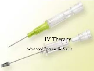
Selecting the IV catheter • Two types of needles and catheters are commonly used in peripheral lines: • Over-the-needle catheters • Winged-tip or scalp-vein needles
IV catheters • The higher the number, the smaller the gauge • The larger the gauge, the more fluid that can be delivered • The shorter the catheter the more fluid that can be delivered
General rule of thumb for selection of size: • Medical patient’s • Use at least a 20 gauge catheter • A 22 gauge can be used if the patient has small, fragile veins • Pediatric patient’s (NOT USED on ages SIX and UNDER) • Use 20-22 gauge • Trauma patient’s • Use at least an 18 gauge catheter