Download
1 / 25
250 likes | 357 Vues
Investigating the primed state of CVD diamond using light and alpha particles to understand the charge collection efficiency with varying bias polarity. The study explores the effects of blue light on electrons and holes, as well as the impact of X-ray priming on charge collection efficiency. Results show improvements in electron response and count rates with blue light. The research also delves into the decay of the primed state, electron collection under white light, and persistent photoconductivity.

E N D
Characterization of primed state of CVD diamond by light and alpha particles C. Manfredotti Experimental Physics Department University of Torino INFN- Sezione di Torino
Erasing previous sample history Experimental set up Priming Investigation of the primed state with light:ALPHA PARTICLE DETECTION Time sequence of measurements
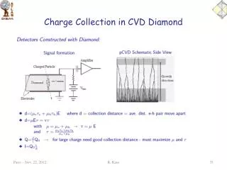
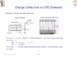
Alpha particle detection can discriminate between hole and electron contribution to Charge Collection Efficiency (CCE) by changing bias polarity and by profiting of the short range of 5.5 MeV alpha particles in diamond (13 mm) Bias voltage (positive) and signal + Electrons cover the longest part of the sample thickness and give the largest contribution to charge signal. e- L Dx Ramo’s theorem: CCE (for one carrier)= Dq/q = Dx/L h+ - Holes almost do not contribute: the range of a particle is too short. a particle
Green, red and infrared light are not able to affect the primed state Blue light enhances electron response and lowers hole response Remember TL? Blue light bleaching is the same and affects holes X-ray priming enhances holes response Charge collection efficiency as a function of priming and of light sensitization
IBIC ( Ion Beam Induced Charge) measurements Effect of light on maps of Charge Collection Efficiency ( CCE )
IBIC maps on De Beers detector primed ( 10 Gy) Response is still better for holes, but counting rate is very low (counting efficiency is 5 – 10%) and detector polarizes completely (no counts) after 1 – 2 hours of irradiation holes electrons Bias – 600 V Bias + 600 V
IBIC maps after priming and during blue light (450 nm) illumination • Counting efficiency improves dramatically, particularly for electrons • Response and uniformity for electrons are much better than for holes. • Charge collection distance (CCD) for electrons can reach 600 mm ! Bias – 600 V Bias + 600 V
Effects of priming and light ( 400 nm ) on alpha spectraTime behaviour of alpha spectraDiscrimination between electrons and holes Preliminary data
Alpha spectra – Electrons Priming – Priming + light From these spectra, time evolution of both centroid and total counts ( integral ) has been derived
Decay of primed state – Different amounts of primingShort termElectrons
Effect of white light on electron collectionTungsten lamp, no interf. filter
Conclusions • Only blue light ( at least below 500 nm ) affects the primed state • Holes : blue light reduces the average CCE of the primed state • Electrons : blue light improves the average CCE • After a short transient ( 1 hr ) both X-ray primed state hole contribution to CCE and blue light continuously primed state electron contribution to CCE seem stable in time ( 30 hr ) • Blue light priming is different from sample to sample and it is independent of amount of previous X-ray priming – even better with no previous priming • Hole reponse ( PC, alpha spectra ) is sensitive to low amounts of priming ( and it is linear with doses up to few tens of mGy )
Priming effects on photoconductivityPPC Persistent PhotoConductivity
Deeper levels are involved Increasing photon energy BGPC increases Increasing number of incident photons More detrapping BGPC decreases Dependence of PC on dose priming dose Sample A Priming fills hole traps. After annealing at 360°C (restoring of the unprimed state), the band at 2.4 eV disappears. It disappears also if the BGPC measurements are carried out starting from higher energy values. Why a maximum at 2.4 eV?
PC peak position and height depend on time and on illumination intensity The sample response depends on its “history”. Annealing: heating at 360 °C for 60 seconds for three times, performed in order to restore the starting conditions. • Factors affecting the BGPC value: • Surface exposed to b-rays (growth or substrate). • Undesired exposure to room light. • Time elapsed before starting the measurements. • Total number of incident photons (exposure time at each wavelength).
Decay of the primed state under illumination For energies of the incident photons between 1.76 and 4.80 eV, the BGPC signal after the priming decreases with a decay that can be fitted using the expression: exp[(-t/τ)β] with β < 1. • Effect of the priming: • filling hole traps. • Decay of the primed state: • optical detrapping of holes
Gain factor Ratio Sprimed / Svirgin The photoconductive gain around 2.0 - 2.1 eV with respect to unprimed case is very high, more than 300 in the case of sample B. This was observed also for other samples, with no evidence of dependence on their electronic quality.
Linearity range Applications in dosimetry Diamond can be used as a passive solid state dosimeter for bio-medical applications. Its attractiveness essentially stems from its radiation hardness, chemical stability against all the body fluids and its absolute nontoxicity. Moreover, diamond is to be considered as a tissue equivalent material since its atomic number is close to the effective atomic number of soft tissue (5.92 for fat and 7.4 for muscle). • Advantages • both large-area detectors • or miniaturised detectors • high sensitivity • good spatial resolution