Tubulointerstitial Disease
Tubulointerstitial Disease. Prof Dr.Gülçin Kantarcı Yeditepe University Nephrology Department. Aims & objectives. State the definition of tubulointerstitial disease. Identify clinical signs of tubulointerstitial disease. Explain the pathophysiology of tubulointerstitial disease.

Tubulointerstitial Disease
E N D
Presentation Transcript
TubulointerstitialDisease Prof Dr.Gülçin Kantarcı Yeditepe UniversityNephrologyDepartment
Aims & objectives • State the definition of tubulointerstitial disease. • Identify clinical signs of tubulointerstitial disease. • Explain the pathophysiology of tubulointerstitial disease.
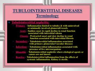
TubulointerstitialNephrithis The tubulointerstitial compartment is affected in all the forms of renal disease. We can findthepathology astubular damage, tubular atrophy, edema, interstitial inflammationor fibrosis. Acute interstitial or tubular damage can produce acute renal failure, Chronicchanges are a good indicator of irreversible lesions primary injury to renal tubules and interstitium resulting decreased renal function.
AcuteTubulointerstitialNephrithis The acute form is most often due to allergic drug reactions or to infections.
suggested by history and urine and blood tests and confirmed by biopsy. Exclusion of glomerular disease Urinalysis: proteinuria<1 g/day (Proteinuria is usually minimal but may reach nephrotic range with combined ATIN-glomerular disease induced by NSAIDs) WBC casts and granular casts Eosinophiluria Low molecular weight proteinuria (LMW-P) ( B2 micglobulin, Tamm-Horsfall gylcoprotein) Diagnosis of ATIN
Tamm-Horsfall gylcoprotein • The most abundant protein of renal origin • Synthesized by cells of the thick ascending limb of the loop of Henle • Excreted in the urine at a relatively constant rate • Urinary excretion can increase following injury to the distal tubule
Diagnosis of ATIN • Eosinophiluria has a positivepredictivevalue of 50% and a negativepredictivevalue of 90% for ATIN; absencesignificantlyexcludesdisease. Blood test findings of tubulardysfunctioninclude; • hyperkalemia (defect in K excretion) • metabolicacidosis(defect in acidexcretion) Ultrasoundfindings:Thekidneysmay be greatlyenlargedandechogenicbyexaminationbecause of interstitialinflammatorycellsandedema.
Renalbiopsy • not often performed for diagnostic purposes but has helped characterize the nature and progression of tubulointerstitial disease. • Glomeruli are usually normal. The earliest finding is interstitial edema, typically followed by interstitial infiltration with lymphocytes, plasma cells, eosinophils, and a few PMNs. • The presence of granulomas suggests sarcoidosis.
Acute tubulointerstitial nephritis (ATIN) • associatedwith an inflammatoryinfiltrateandedemainvolvingtherenalinterstitium (daystomonths). • Over 95% of casesresultfrominfectionor an allergicdrugreaction; • a syndrome of ATIN associatedwithuveitis (renal-ocular syndrome-TINU) alsooccursand is idiopathic.
causes Acute Kidney Injury (AKI) • severe cases, delayed therapy, or continuance of an offending drug can lead to permanent injury with Chronic Renal Failure (CRF)
Symptoms and signs of ATIN • Fever • urticarial rash • Onset may be as long as several weeks after a 1st toxic exposure or as soon as 3 to 5 days after a 2nd exposure; • extremes in latency range from 1 day with rifampin to 18 mo with an NSAID.
Symptoms and signs of ATIN • Abdominal pain, weight loss, and bilateral renal masses (caused by interstitial edema) may also occur and with fever may mistakenly suggest renal malignancy or polycystic kidney disease. • Many patients develop polyuria and nocturia (defect in concentration and Na reabsorption). • Peripheral edema and hypertension are uncommon unless renal insufficiency or renal failure occurs
Maculopapular rash in a patient with drug–induced acute interstitial nephritis
On lightmicroscopy, interstitialinfiltrationwithmononuclearcells, with normal glomeruli. It is usuallyassociatedwithinterstitialedema, andwithtubularlesions
prognosis • vary by the etiology and potential reversibility of the disorder at the time of diagnosis.
Prognosis • In drug-induced ATIN, renal function usually recovers within 6 to 8 wk when the offending drug is withdrawn, although some residual scarring is common. • Recovery may be incomplete, with persistent azotemia above baseline
histologic changes usually are reversible if the cause is recognized and removed; however, some severe cases progress to fibrosis and renal failure. • Regardless of cause, diffuse rather than patchy interstitial infiltrates, delayed response to prednisone , and persistent acute renal failure (> 3 wk) suggest irreversible injury.
Chronictubulointerstitialnephritis The CTIN is irreversible and rather than allergic condition, it is associated with genetic metabolic obstructive uropathy chronic environmental toxins drugs and herbs.
Chronictubulointerstitialnephritis (CTIN) • chronic tubular insults cause gradual interstitial infiltration and fibrosis, tubular atrophy and dysfunction, and a gradual deterioration of renal function, usually over years. • Glomerular involvement (glomerulosclerosis) is much more common in CTIN than in ATIN.
Causes of CTIN • infections, • reflux or obstructive nephropathy, • drugs, and • other diseases. • toxins, • metabolic diseases, • hypertension • inherited disorders results in symmetric and bilateral disease;
Diagnosis of CTIN • Findings of CTIN are generally similar to those of ATIN, though urinary RBCs and WBCs are uncommon. Because CTIN is insidious in onset and is associated with interstitial fibrosis, imaging tests may show small kidneys with evidence of scarring and asymmetry.
Pathogenesis of tubulointerstitialfibrosis Injury to the tubule and peritubular capillary leads to the • generation of chemotactic and adhesive factors that result in macrophage and T–cell accumulation.
Local macrophage and fibroblast activation ensues, driven by growth factors such as PDGF(platelet–derived growth factor) and TGF-β (transforming growth factor- β), which results in collagen production by tubular cells and fibroblasts,eventually culminating in the fibrotic lesion.
Renal biyopsy • In CTIN, glomeruli vary from normal to completely destroyed. • The interstitium contains varying degrees of inflammatory cells and fibrosis.
Tubular atrophy • Tubules may be absent or atrophied • Nonscarred areas appear almost normal. Grossly, the kidneys are small and atrophic
Tubular casts. Casts are periodic acid–Schiff stain (PAS) positive and usually contain Tamm–Horsfall protein. Some may contain desquamated tubular cells and macrophages. (Periodic acid–Schiff ×400).
An intravenousurogram of a patientwithanalgesicnephropathy • The pyelogram shows evidence of a necrotic papilla in the lateral minor calyx producing a 'ring' sign
Prognosis of CTIN • depends on the cause and on the ability to recognize and stop the process before irreversible fibrosis occurs. • Many genetic (cystic kidney disease), metabolic (cystinosis), and toxic (heavy metal) causes may not be modifiable, in which case CTIN usually evolves to end-stage renal disease
ANALGESIC ABUSE NEPHROPATHY • Analgesic abuse nephropathy (AAN) is CTIN caused by cumulative lifetime use of large amounts (eg, ≥ 2 kg) of certain analgesics. • AAN predominates in women (peak incidence, 50 to 55 yr) and, in the US, is responsible for 3 to 5% of cases of end-stage renal disease (13 to 20% in Australia and South Africa).
AAN was originally described in conjunction with overuse of combination analgesics containing phenacetin (typically with aspirin, acetaminophen, codeine, or caffeine).
Patientspresentwith; • renalinsufficiency, • a blandurinarysediment, • non-nephroticproteinuria. • Hypertensionand • impairedurinaryconcentrationarecommon.
Flank pain and hematuria are signs of papillary necrosis that occur late in the course of disease. • Chronic complaints of musculoskeletal pain, headache, malaise, and dyspepsia may be precipitants of long-term analgesic use rather than effects of AAN.
Renal function stabilizes when analgesics are stopped unless renal insufficiency is advanced, in which case it may progress to renal failure. • Patients with AAN are at greater risk of transitional cell carcinomas of the urinary tract.
METABOLIC NEPHROPATHIES Acuteuratenephropathy:This is not a true form of ATIN but rather an intraluminalobstructiveuropathycausedbyuricacidcrystaldepositionwithinthelumen of renaltubules; acuteoliguricoranuricrenalfailureresults. • Itmostcommonlyoccursfromtumorlysissyndrome
Chronic urate nephropathy:This condition is CTIN caused by deposition of Na urate crystals in the medullary interstitium in the setting of chronic hyperuricemia. • Chronic urate nephropathy was once common in patients with tophaceous gout but is now rare because of treatment
Hyperoxaluria • a cause of both acute and chronic tubulointerstitial nephritis. • ATIN and AKI may develop in susceptible patients who ingest high-oxalate foods (eg, tea, chocolate, spinach, star fruit) or who are exposed to exogenous substances that are metabolized into oxalate (eg, ethylene glycol ingestion, methoxyflurane anesthesia, large doses of ascorbic acid).
CTIN and progressive chronic renal failure develop in patients with inherited disorders of excessive oxalate production (types I and II primary hyperoxaluria) or acquired GI diseases (eg, short bowel syndrome with increased gut absorption).
Symptoms and signs • Differ by form of disease and include; • hematuria • renal colic from oxalate calculi, • UTI and pyuria, • hypertension, • renal tubular acidosis
HEAVY METAL NEPHROPATHY • Lead: accumulates in proximal tubular cells. Chronic lead exposure (5 to 30 yr) causes progressive tubular atrophy, interstitial fibrosis, hypertension, and gout. Chronic low-level exposure may cause renal insufficiency and hypertension independent of tubulointerstitial disease. Children exposed to lead paint dust or chips, battery workers, and drinkers of moonshine alcohol are most at risk.
Diagnosis • usually made by whole blood lead levels. Alternatively, x-ray fluorescence
HEAVY METAL NEPHROPATHY • Cadmium:Cadmium from contaminated water, food, and tobacco and from workplace exposures can cause nephropathy. • Diagnosis • a history of occupational exposure, • increased levels of urinary β2-microglobulin, • increased urinary cadmium levels (> 7 μg/g creatinine).
HEAVY METAL NEPHROPATHY • Other heavy metals:Those that are nephrotoxic include copper, gold, uranium, arsenic, iron, mercury, bismuth, and chromium. • All cause tubular damage and dysfunction but glomerulopathies may predominate with some compounds (mercury, gold). • Treatment involves removal of the patient from further exposure and chelating agents (copper, arsenic, bismuth) or dialysis (chromium, arsenic, bismuth).
REFLUX NEPHROPATHY • Refluxnephropathy is renalscarringinducedbyvesicoureteralreflux of infectedurineintotherenalparenchyma. • Chronicpyelonephritisalsomayplay a role, but UTI withoutintrarenalreflux is unlikelytocausenephropathy.