Download
1 / 26
420 likes | 2.34k Vues
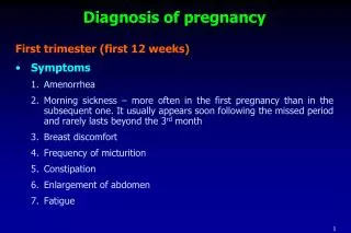
Diagnosis of pregnancy. First trimester (first 12 weeks) Symptoms 1. Amenorrhea 2. Morning sickness – more often in the first pregnancy than in the subsequent one. It usually appears soon following the missed period and rarely lasts beyond the 3 rd month Breast discomfort

E N D
Diagnosis of pregnancy First trimester (first 12 weeks) • Symptoms 1. Amenorrhea 2. Morning sickness – more often in the first pregnancy than in the subsequent one. It usually appears soon following the missed period and rarely lasts beyond the 3rd month • Breast discomfort • Frequency of micturition • Constipation • Enlargement of abdomen • Fatigue
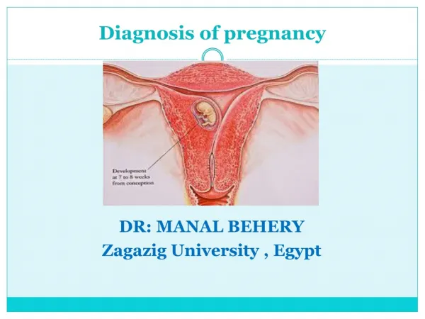
Diagnosis of pregnancy (contd…) • Signs • Breast: Engorgement of breast with dilatation of superficial veins Areola more pigmented Montgomery’s tubercles are prominent Secretion as early as 8th week • Per abdomen – uterus remains a pelvic organ until at 12th week • Pelvic changes • Chadwick’s sign – It is the dusky blue of anterior vaginal wall visible at about 8th week of pregnancy. The discoloration is due to local vascularity. • Uterine signs – the pregnant uterus feels soft and elastic • Hegar’s sign – 6-10 weeks
Diagnosis of pregnancy (contd…) • Immunological test • Depends on agglutination reaction of the antigen (b-HCG) • Sonography • Gestational ring • Cardiac motion uniformly by 7th week • Second trimester (13-28 weeks) • Quickening • Progressive enlargement of the lower abdomen • General examination • Chloasma
Diagnosis of pregnancy (contd…) • Abdominal examination • Inspection • Fundal height • Palpation • The uterus feels soft and elastic • Braxton-Hicks contractions • Palpation of fetal parts • Active fetal movements • Auscultation • Last trimester (29-40 weeks) • Enlargement of the abdomen • Fetal movement • Braxton-Hicks • Fetal movements
Physiological changes during pregnancy • Vulva • Superficial varicosities may appear • Labia minora are pigmented and hypertrophied • Vagina • Increased blood supply of the venous plexus surrounding the walls give the bluish coloration of the mucosa (Jacquemier’s sign) • Secretion • The secretion becomes copious • pH becomes acidic (3.5-6) • Uterus • The uterus which is non-pregnant state weighs about 50mg and measures about 7.5 cm in length, at term weighs 1000 gms and measures 35 cm in length. Hypertrophy and hyperplasia of the cells.
Physiological changes during pregnancy (contd…) Cutaneous changes • Face • Chloasma gravidarum or pregnancy mask, extreme form of pigmentation around the cheek, forehead and around the eyes. It may be patchy or diffuse, disappears spontaneously after delivery • Abdomen • Linea nigra – brownish black pigmented area in the midline stretching from the xiphisternum to the symphysis pubis • Striae gravidarum – represent the tissue in the deeper layer of the cutis, mechanical stretching • Weight gain • The total weight gain during the course of a singleton pregnancy in average is 10kgs.
Cardiovascular changes (1) • Position and size of heart • ECG changes Increased heart rate (+15%) 15-degrees left axis deviation Inverted T-waves in lead III Q in lead III and AVF Unspecific ST changes • Appears larger on X-ray
Cardiovascular changes (2) • Murmurs: soft , transient • Inferior vena cava syndrome: In the supine position, the inferior vena cava is compressed by the enlarged uterus, resulting in decreased cardiac output. Some women may have symptoms that include dizziness, light-headedness, and syncope.
Cardiovascular changes (3) • Stroke volume +30% • Heart rate +15% • Cardiac output +40% • Oxygen consumption +20% • SVR (systemic vascular resistance)-5% • Systolic BP -10mmHg • Diastolic BP -15mmHg • Mean BP -15mmHg
Cardiovascular changes (4) • Blood volume +30% • Plasma volume +40% • Red blood cell volume +20%
Cardiovascular changes(5) • Venous pressure: • unchanged in the upper body • Significantly increases in the lower extremities, esp. during supine, sitting or standing position, returns to near normal in lateral recumbent position
Hematologic system (1) • Blood volume (polymorphonuclear)+40% • Dilutional anemia Hb 110 g/L • Leukocytosis 15,000/ml • Platelet no change • Sedimentation rate increase, 100m/h
Hematologic system (2) • Clotting factors: hypercoagulable, throboembolism Fibrinogen (factor I) +50% (4.5 vs 3 g/L) Factor VIII increase Factors VII, IX, X and XII increase Prothrombin time, PT shortened ATPP activated partial thromoplastin time shortened Fibrinolytic activity decrease
Hematologic system (3) • Iron : active transplacental transfer Requirement 1000mg increase maternal red cell mass 500mg fetal development 300mg compensate for normal iron loss 200mg
Physiological changes during pregnancy (contd…) • Leucocytes • Neutrophilic leucocytosis occurs to the extent of 10-15,000/cu.mm and even to 20,000/cu.mm • Total proteins • Supine hypotension syndrome (postural hypotension) • Carbohydrate metabolism • Glycosuria – lowered renal threshold, increased glomerular filtration rate.
Physiological changes during pregnancy (contd…) Systemic changes • Nervous system • May be generalized neuritis probably due to vitamin B1 deficiency • Compression of the lumbosacral trunk by the fetal head or by features of sciatica • Compression of the median nerve (Carpal tunnel syndrome)
Hemodynamic changes during pregnancy • Decreased peripheral vascular resistance • Decreased pulmonary vascular resistance • Decreased colloid oncotic pressure • Increased cardiac output • Increased pulse rate
Changes in the kidneys and the urinary system Anatomical changes • Dilatation of the collecting system • The renal calices, the renal pelvis and the ureters starts to dilate and remain enlarged for several weeks after delivery • Causes • Progesterone • Compression of the ureter Physiological changes • The most important consequence of the increased RPF is a 50% increase in the GFR • The serum creatinine and urea nitrogen concentration below lower than in the non-pregnant situation
Endocrinology in relation to reproduction • Hormones of placenta • Protein hormones • Human chorionic gonadotrophin (HCG) • Human placenta lactogen (HPL) • Human chorionic thyrotrophin (HCT) • Human chorionic corticotrophin (HCC) • Pregnancy specific b-1 glycoprotein (PS b G) • Steroid hormones • Estrogens – estriol, estradiol and estrone • Progesterone
Endocrinology in relation to reproduction (contd…) • Estrogen and progesterone • 100 folds increase in progesterone concentration • Estrogen levels are also very high • The level of SHBG increases • Adrenal cortical hormones • Increased level of plasma cortisol • There is increase in adrenal androgens – helps in protein anabolism • Increase in aldosterone secretion
Endocrinology in relation to reproduction (contd…) • Thyroid gland • Moderate enlargement with hyperplasia • Increased secretion of thyroid hormones • Parathyroid gland • Enlarged with increase secretion of parathyroid hormone to facilitate mobilization of ionic calcium and phosphorus for fetal bone development • Calcitonin level slightly increased just to counter the effect of PTH on maternal skeleton • Pancreas • Hypertrophy and hyperplasia of beta cells of Langerhans • Pregnancy has diabetogenic effect
Endocrinology in relation to reproduction (contd…) Diabetogenic effects of pregnancy Insulin resistance • Production of placental somatomammotropin • Increased production of cortisol, estriol, and progesterone • Increased insulin destruction by kidney and placenta
Endocrinology in relation to reproduction (contd…) • Human chorionic gonadotrophin (HCG) • Functions • Secretion of progesterone by the corpus luteum of pregnancy • HCG stimulates Leydig cells of the male fetus to produce testosterone in conjunction with fetal pituitary gonadotropins. It is thus indirectly involved in the development of male external genitalia • It has got immuno-suppressive activity which may inhibit the maternal processes of immunorejection of the fetus as a homograft
Endocrinology in relation to reproduction (contd…) Steroidal hormones • Functions • Estrogen causes hypertrophy and hyperplasia of the uterine myometrium, thereby increasing the accommodation capacity, vascularity and blood flow of the uterus. • Progesterone in conjunction with estrogen stimulates growth of the uterus • Development and hypertrophy of the breasts. Hypertrophy and proliferation of the ducts are due to estrogen • Both the steroids are required for the adaptation of the maternal organs to the constantly increasing demands of the growing fetus • The steroids are involved in the complex pathway in initiation of normal labor
Metabolic changes • Carbohydrate metabolism • Fetus drives its energy almost totally from glucose, passed through placenta by facilitated diffusion • Lipid metabolism • Increased mobilization of lipids from maternal adipose tissue to raise plasma FFA level • HPL has glucose sparing effect by mobilizing free fatty acids for mothers skeletal and cardiac muscles and diverting the glucose to placenta and fetus
Metabolic changes (contd…) Salt and water metabolism • Marked water retention is found in pregnancy with the decrease in plasma osmolarity • Edema of legs seen because of increased venous pressure due to compression by gravid uterus • Increase in blood volume causes decreased oncotic pressure causes leakage of water in the tissue bed. • The reduction in serum, sodium is caused by increased GFR. However, sodium and fluid balance is maintained by increase in plasma aldosterone and increase level of estrogen and deoxycorticosterone prevents sodium loss