Download

1 / 63
1.36k likes | 2.32k Vues
Diagnosis of Pregnancy. Shanghai OB/GYN Hospital Fudan University Yuan Lu. LMP: Last normal menstrual period Gestational age 280 days 40 weeks. Duration of Pregnancy. Estimation of Gestation Age & Prediction of Excepted Date of Delivery. Excepted Due Date (EDD)

E N D
Diagnosis of Pregnancy Shanghai OB/GYN Hospital Fudan University Yuan Lu
LMP: Last normal menstrual period • Gestational age • 280 days • 40 weeks Duration of Pregnancy
Estimation of Gestation Age& Prediction of Excepted Date of Delivery • Excepted Due Date (EDD) = LMP﹣3/﹢9 month and ﹢7 days LMP: Sep 10, EDD: June 17.
First trimester: first 12 weeks • Second trimester: 13-28 weeks • Last trimester: 29-40 weeks
First trimester • Symptoms • Signs • Immunological Tests • Ultra Sonograph Pregnancy Diagnosis
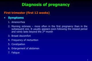
Symptoms Amenorrhoea Morning Sickness Frequence of micturition Breast discomfort Fatigue
Symptoms • Amenorrhoea cardinal/most important sign of early pregnancy One or two episodes of bloody discharge
Morning Sickness Usually appears soon following the missed period; Rarely lasts beyond 3 months
Frequence of micturition • troublesome symptom during 8-12 weeks • enlarged uterus • congestion of bladder • increased intravascular volume
Breast discomfort • Fullness • Pricking sensation
Fatigue Occur early in pregnancy
Signs • Breast changes • valuable only in primigravidae • breast changes are evident between 6-8 weeks • vascular engorgement & nipple and areola pigment • colostrum expressed as early as 12th weeks
Uterus Fundus Height • Uterine sign • Uterus remains a pelvis organ until 12 weeks • Size, shape and consistency 6th week 8th week 12th week
Medical Signs Non pregnant uterus Pregnant uterus Chadwick’s sign: darkening of the cervix, vagina, and vulva; Goodell’s sign: softening of the vaginal portion of the cervix Hegar’s sign: softening of the uters isthmus; Linea nigra: pigmentation of linea alba The pregnant uterus feels softand elastic
human chorionic gonadotropin (hCG): α/β-subunit produced in the syncytiotrophoblast Urine approximately 4 weeks following the first day of the last menstrual period All urine pregnancy tests are best performed on early-morning urine specimens, which contain the highest concentration of hCG Serum specific and sensitive by following serial quantitative hCG levels and comparing them to the expected rise derived from normative data for proven normal intrauterine pregnancies Pregnancy test
the hCG usually doubles every 48-72 HOURS it normally increases by at least 60% every 2 DAYS
Ultrasound examination • Abdominal ultrasound: allowing visualization of a normal pregnancy gestational sac 5 to 6 weeks after the beginning of the last normal menstrual period (corresponding to β-hCG concentrations of 5000 to 6000 mIU/mL) • Transvaginal ultrasound: often detects pregnancy at 3 to 4 weeks of gestation (corresponding to β-hCG concentrations of 1000 to 2000 mIU/mL)
Image of an early gestational sac containing a yolk sac and early embryo. The yolk sac is the circular hyperechoic structure adjacent to the embryo. Image of an early gestational sac demonstrating the early embryo. Calipers are placed at both ends of the embryo measuring the longest length from the "crown to the rump" giving the crown-rump length. This measurement is used for dating the pregnancy.
First Trimester Review The Whole Period of Pregnancy Can Be Divided Into Three Stages • The first trimester (early pregnancy): 1-12w • The second trimester (middle pregnancy): 13-27 w • The third trimester (late pregnancy): 28-40w
1. History and symptoms • Cessation of menstruation • Nausea and Vomiting • Urinary symptoms • Mastodynia
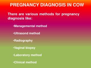
2. Signs • Breast changes • Changes of the reproductive organs (Vagina, Cervix, Uterus) 3. Supplementary examination • Pregnancy test • Basal body temperature (BBT) • Progesterone test • Ultrasonography
Symptoms • Abdominal examination • Vaginal examination
Symptoms • Nausea, vomiting, frequency of micturition subside • Amenorrhea continues • Quickening: perception of active fetal movement by women (From 18th week) • Progressive enlargement of lower abdomen by the growing uterus
Abdominal examination Inspection Palpation Auscultation
Inspection • Linea nigra • Striae Ensiform Cartilage Symphysis Pubis Striae
Palpation • Fundal height increases • Uterus soft and elastic, ovoid in shape • Braxton-Hicks Contraction • Palpation of fetal parts: 20th week • Active fetal movements: 20th week • External ballottement
Fundal height • 16th week: midway between symphysis pubis and umbilicus • 22~24th week: at the level of umbilicus • 28th week: at the junction of the lower 1/3 and upper 2/3 of the distance between the umbilicus and ensiform cartilage
Abnormal Fundal Height • IUGR (intrauterine growth retardation) • Multiple Pregnancy • Polyhydramnios(CNS or Cardiovascular Disfunction) • Oligohydramnios
Braxton-Hicks Contraction • Cause • a tightening of the uterine muscles for one to two hours and are thought to be an aid to the body in its preparation for birth. • Alleviating factors • Rhythmic breathing • Lying down on the left side • A slight change in movement • Urination
Very early, the uterus undergoes spontaneous contraction • Firmer at one moment and soft at another • Can be excited by rubbing the uterus • Irregular, infrequent, spasmodic, and painless • Near term, frequent with increase in intensity, discomfort • Merge with the labor
Palpation of fetal parts Diagnosis of pregnancy Identify the presentation and position of fetus
Active fetal movements Positive evidence of pregnancy & live fetus Faint flutter→stronger movement
Ascutation • Fetal heart sound • Most conclusive • 18-20 weeks • Location • 140-160 bpm→120-140 bpm
Vaginal Examination • The bluish discolouration of the vagina, cervix is much more evident; • Cervix softening
Ultrasound examination • Sonograph: 12-20 weeks; a detailed survey of fetal anatomy, placenta localization, integrity of the cervical canal
Second Trimester Review • Symptoms • Abdominal enlargement and fetal movement generally occurs after the 18th to 20th week of gestation.
Signs • The uterus continues to enlarge • Fetal movement (quickening) can usually be seen or heard after 18th week of gestation • Fetal heart sound can be heard at rate varies from 120 to 160 beats per minute. • The fetal body can usually be palpated by the 18th to 20th week of gestation unless the patient is too fat, the abdomen is tender or there is an excessive amount of amniotic fluid.
Symptoms • Amenorrhoea • Enlargement of the abdomen • Lightening: due to the engagement of the presenting part • Frequency of micturition • Fetal movement
Sign • Cutaneous changes: increased pigmentation and striae • Uterine shape: cylindrical to spherical beyond 36th week • Fundal height • Braxton-Hicks contraction • Fetal movement • Palpation of the fetal parts
32th week: the junction of the upper and middle third between the distance of umbilicus and ensiform cartilage 36th week: the level of the ensiform cartilage 40th week: down to the level of 32th
Symphysisfundal height (SFH) • After 24 weeks, the SFH measured in cm. correspond to the number of the weeks up to 36 weeks. • A variation of ± 2 is accepted as normal. • Variation beyond the normal range needs further evaluation.
Four steps of obstetric palpation Upper part of the uterus Lateral part of the uterus Presentation; Engagement Further confirmation
The first step-Fundal grip broad, soft, irregular mass- head smooth, hard, globular – breech The second step smooth curved and resistant-black comparatively empty and small knob -limb Four steps of obstetric palpation
The third step confirm the present The fourth step determine its engagement degree Four steps of obstetric palpation