Innate Immunity
700 likes | 760 Vues
Innate Immunity. 15. An Overview of the Body ' s Defenses. Resistance to most plant and animal pathogens Species resistance Due to physiological processes of humans that are incompatible with those of the pathogen Correct chemical receptors are not present on human cells

Innate Immunity
E N D
Presentation Transcript
An Overview of the Body's Defenses • Resistance to most plant and animal pathogens • Species resistance • Due to physiological processes of humans that are incompatible with those of the pathogen • Correct chemical receptors are not present on human cells • Conditions may be incompatible with those needed for pathogen's survival • Humans do not have innate resistance to a number of pathogens
An Overview of the Body's Defenses • Three main lines of defense • Innate immunity — first two lines of defense • External physical barriers to pathogens • Protective cells, bloodborne chemicals, and processes • Adaptive immunity
An Overview of the Body's Defenses • Tell Me Why • Why aren't the body's skin and mucous membrane barriers significant factors in your resistance to infection by hyperthermophiles?
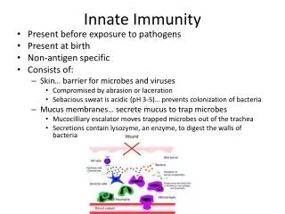
The Body's First Line of Defense • Structures, chemicals, and processes that work to prevent pathogens entering the body • Skin and mucous membranes of the respiratory, digestive, urinary, and reproductive systems
The Body's First Line of Defense • The Role of Skin in Innate Immunity • Skin is composed of two major layers • Epidermis • Multiple layers of tightly packed cells • Few pathogens can penetrate these layers • Shedding of dead skin cells removes microorganisms • Epidermal dendritic cells phagocytize pathogens • Dermis • Collagen fibers help skin resist abrasions that could introduce microorganisms
Figure 15.1 A scanning electron micrograph of the surface of human skin.
The Body's First Line of Defense • The Role of Skin in Innate Immunity • Skin has chemicals that defend against pathogens • Antimicrobial peptides (defensins) secreted by dermal cells • Perspiration secreted by sweat glands • Salt inhibits growth of pathogens • Antimicrobial peptides called dermcidins act against many bacteria and fungi • Lysozyme destroys cell wall of bacteria
The Body's First Line of Defense • The Role of Skin in Innate Immunity • Skin has chemicals that defend against pathogens • Sebum secreted by sebaceous (oil) glands • Helps keep skin pliable and less likely to break or tear • Lowers skin pH to a level inhibitory to many bacteria
The Body's First Line of Defense • The Role of Mucous Membranes in Innate Immunity • Mucous membranes line all body cavities open to environment • Two distinct layers • Epithelium • Thin, outer covering of the mucous membranes • Epithelial cells are living • Tightly packed to prevent entry of many pathogens • Continual shedding of cells carries away microorganisms • Dendritic cells below epithelium phagocytize pathogens • Goblet and ciliated columnar cells help remove invaders • Deeper connective layer that supports the epithelium • Produce chemicals that defend against pathogens
Figure 15.2 The structure of the respiratory system, which is lined with a mucous membrane. Nasal cavity Pharynx Tongue Epiglottis Larynx (voice box) Esophagus Trachea Bronchus Bronchioles
The Body's First Line of Defense • The Role of the Lacrimal Apparatus in Innate Immunity • Lacrimal apparatus • Produces and drains tears • Blinking spreads tears and washes surface of the eye • Lysozyme in tears destroys bacteria
Figure 15.3 The lacrimal apparatus. Nasolacrimal duct Paranasal sinus Lacrimal apparatus Lacrimal glands Pharynx Lacrimal gland duct Lacrimal canal Nasolacrimal duct Trachea Esophagus Lateral view Anterior view
The Body's First Line of Defense • The Role of Normal Microbiota in Innate Immunity • Microbial antagonism • Normal microbiota compete with potential pathogens • Activities of normal microbiota make it hard for pathogens to compete • Consume nutrients • Create an environment unfavorable to other microorganisms • Help stimulate the body's second line of defense • Promote overall health by providing vitamins to host
The Body's First Line of Defense • Other First-Line Defenses • Antimicrobial peptides • Present in skin, mucous membranes, neutrophils • Act against a variety of microbes • Work in several ways • Other processes and chemicals • Many organs secrete chemicals with antimicrobial properties
The Body's First Line of Defense • Tell Me Why • Some strains of Staphylococcus aureus produce exfoliative toxin, a chemical that causes portions of the entire outer layer of the skin to be sloughed off in a disease called scalded skin syndrome. Given that cells of the outer layer are going to fall off anyway, why is this disease dangerous?
The Body's Second Line of Defense • Operates when pathogens penetrate the skin or mucous membranes • Composed of cells, antimicrobial chemicals, and processes • Many of these components are contained or originate in the blood
The Body's Second Line of Defense • Defense Components of Blood • Plasma • Mostly water containing electrolytes, dissolved gases, nutrients, and proteins • Serum is the fluid remaining when clotting factors are removed • Contains iron-binding compounds • Iron is needed for metabolism • Some microbes produce iron-binding proteins (siderophores) • Complement proteins and antibodies are also found in plasma
The Body's Second Line of Defense • Defense Components of Blood • Defensive blood cells: leukocytes • Cells and cell fragments in plasma are called formed elements • Three types of formed elements • Erythrocytes • Carry oxygen and carbon dioxide in the blood • Platelets • Involved in blood clotting • Leukocytes • Involved in defending the body against invaders • Classified as granulocytes and agranulocytes
Figure 15.4 A schematic representation of hematopoiesis. Blood stem cell in bone marrow Erythroid stem cell Myeloid stem cell Lymphoid stem cell Erythrocyte Lymphocyte Basophil Platelets Neutrophil Monocyte Eosinophil Inflammation Phagocytosis Clotting, inflammation Innate immunity, second line of defense Adaptive immunity Gas transportation Leukocytes
The Body's Second Line of Defense • Defense Components of Blood • Defensive blood cells: leukocytes • Granulocytes • Contain large granules that stain different colors • Three types • Basophils – stain blue with basic dye methylene blue • Eosinophils – stain red/orange with acidic dye eosin • Neutrophils – stain lilac with mix of acidic and basic dyes • Neutrophils and eosinophils • Phagocytize pathogens • Capable of diapedesis
The Body's Second Line of Defense • Defense Components of Blood • Defensive blood cells: leukocytes • Agranulocytes • Cytoplasm appears uniform under a light microscope • Two types • Lymphocytes • Most involved in adaptive immunity • Natural killer lymphocytes • Monocytes • Leave the blood and mature into macrophages • Phagocytic cells that devour foreign objects
Figure 15.5 Leukocytes as seen in stained blood smears. Basophil 0.5–1% Granulocytes Lymphocyte 20–25% Eosinophil 2–4% Agranulocytes Neutrophil 60–70% Monocyte 3–8%
The Body's Second Line of Defense • Defense Components of Blood • Defensive blood cells: leukocytes • Lab analysis of leukocytes • Differential white blood cell count can signal disease • Increased eosinophils indicate allergies or parasitic worm infection • Bacterial diseases often show increase in leukocytes and neutrophils • Viral infections show increase in lymphocytes
The Body's Second Line of Defense • Phagocytosis • Cells capable of phagocytosis are called phagocytes • Phagocytosis is not completely understood • Can be divided into six stages • Chemotaxis • Adherence • Ingestion • Maturation • Killing • Elimination
Figure 15.6 The events in phagocytosis. Chemotaxis of phagocyte to microbes 1 Neisseria (microbes) Adherence 2 Pseudopodia move (chemotaxis) Ingestion of microbes by phagocytes 3 4 Fusion of a series of vesicles, including lysosomes Phagosome Golgi body Killing of microbes by enzymes and other chemicals 5 Lysosome Nucleus Phagolysosome Residual body Pseudopod 6 Elimination by exocytosis Phagocyte
The Body's Second Line of Defense • Nonphagocytic Killing • Killing by eosinophils • Attack parasitic helminths by attaching to their surface • Secrete toxins that weaken or kill the helminth • Eosinophilia is often indicative of a helminth infestation or allergies • Eosinophil mitochondrial DNA and proteins form structure that kills some bacteria
The Body's Second Line of Defense • Nonphagocytic Killing • Killing by natural killer lymphocytes • Secrete toxins onto surface of virally infected cells and tumors • Differentiate normal body cells because they have membrane proteins similar to the NK cells
The Body's Second Line of Defense • Nonphagocytic Killing • Killing by neutrophils • Can destroy microbes without phagocytosis • Produce chemicals that kill nearby invaders • Generate extracellular fibers called neutrophil extracellulartraps (NETs) that bind to and kill bacteria
The Body's Second Line of Defense • Nonspecific Chemical Defenses Against Pathogens • Toll-like receptors (TLRs) • Integral membrane proteins produced by phagocytic cells • Bind pathogen-associated molecular patterns (PAMPs) • Initiate defensive responses • Apoptosis • Secretion of inflammatory mediators • Stimulate adaptive immune response
The Body's Second Line of Defense • Nonspecific Chemical Defenses Against Pathogens • NOD proteins • Cytosolic proteins that bind PAMPs • Trigger inflammation, apoptosis, and other innate responses • Mechanism of action still being researched • Mutations in NOD genes associated with some inflammatory bowel diseases
The Body's Second Line of Defense • Nonspecific Chemical Defenses Against Pathogens • Interferons • Protein molecules released by host cells to nonspecifically inhibit the spread of viral infections • Cause many symptoms associated with viral infections • Two types • Types I (alpha and beta) • Type II (gamma)
Figure 15.7 The actions of alpha and beta interferons. 1 Virus infects cell. Virus Double- stranded RNA Viral replication in cell triggers transcription and translation of IFN-α or IFN-β, depending on type of host cell. 2 IFN gene Time passes Meanwhile, the infected cell dies, releasing viruses. 5 Nucleus mRNA IFN Infected cell Infected cell at a later time Interferon is released, diffuses to neighboring uninfected cells, and binds to receptors. 3 Interferon receptor When the second cell becomes infected with viruses, double- stranded RNA of the virus activates AVP. 6 Inactive AVP AVP gene Double- stranded viral RNA Active AVPs Binding triggers transcription and translation of inactive antiviral proteins (AVPs). 4 Time passes mRNA Active AVPs degrade mRNA and bind to ribosomes, which stops protein synthesis and viral replication. 7 Ribosome Inactive AVPs mRNA Uninfected neighboring cell Same neighboring cell now protected at the later time
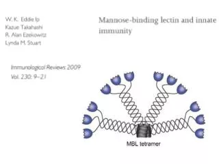
The Body's Second Line of Defense • Nonspecific Chemical Defenses Against Pathogens • Complement • Set of serum proteins designated numerically according to their order of discovery • Complement activation results in lysis of the foreign cell • Indirectly trigger inflammation and fever • Complement can be activated in three ways • Classical pathway • Alternative pathway • Lectin pathway
Figure 15.8 Pathways by which complement is activated. Classical pathway Alternative pathway Lectin pathway Mannose C3b Antigen Endotoxin and glycoproteins Lectins Antibody Factors B, D, and P C3b Complement proteins 1, 2, 4 Complement cascade Activation (C3 C3a + C3b) Opsonization Inflammation C5 convertase C5 C5a + C5b Inflammation Membrane attack complex and cell lysis
Figure 15.9 The classical pathway and complement cascade. H2O Cytoplasmic membrane Membrane attack complex H2O C9 C9 C9 C9 C9 H2O H2O C9 C9 C9 C9 C9 C7 C8 C6 C5b Pathogen C5b combines with C6, C7, C8, and several molecules of C9 to form a membrane attack complex (MAC). A MAC drills a circular hole in the pathogen’s cytoplasmic membrane, leading to lysis of the cell. 6 C1 Causes chemotaxis of phagocytes and triggers inflammation Antigen Antibody C5a C1 becomes an active enzyme when it binds to antibody-antigen complexes. 1 C5b Causes inflammation 5 This enzyme cleaves C5 into C5a and C5b. Enzymatic C1 C4a C4b C4b C3b C2b Enzyme C1 splits molecules of C2 and of C4. 2 Enzyme (C5 convertase) C4 C2a C5 C2b C2 C2b C2 C3b combines with the remaining fragments of C2 and C4 to form a third enzyme. 4 C4a C4 C4b 3 Fragments of C2 and C4 combine to form a second enzyme that splits C3 into C3a and C3b. C2a C4b Enzyme C3b C3a C3 Causes chemotaxis of phagocytes and triggers inflammation C3 Acts as opsonin C3b C3a
Figure 15.10 Membrane attack complexes. Membrane attack complex
The Body's Second Line of Defense • Nonspecific Chemical Defenses Against Pathogens • Complement • Inactivation of complement • Body's own cells withstand complement cascade • Proteins on many cells bind and break down activated complement proteins
The Body's Second Line of Defense • Inflammation • Nonspecific response to tissue damage from various causes • Characterized by redness, heat, swelling, and pain • Two types • Acute • Chronic