Chapter 10 Nucleic Acids and Protein synthesis
180 likes | 309 Vues
This chapter covers the essential roles of DNA and RNA in genetic information processing. DNA (Deoxyribonucleic Acid) stores and transmits genetic information, consisting of nucleotides formed by a sugar, phosphate, and nitrogen base (Adenine, Guanine, Cytosine, Thymine). The double helix structure was proposed by Watson and Crick, with contributions from Franklin and Wilkins. RNA (Ribonucleic Acid), which is single-stranded, moves genetic information from the nucleus for protein synthesis. The processes of transcription and replication are crucial for the accurate copying and expression of genes.

Chapter 10 Nucleic Acids and Protein synthesis
E N D
Presentation Transcript
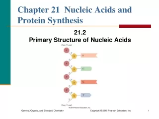
DNA = DeoxyriboNucleic Acid – stores and transmits the genetic information that tells cells which proteins to make and when to make them. • DNA is made up of 2 long chains of NUCLEOTIDES. • Nucleotide = Sugar (deoxyribose) + Phosphate + Nitrogen Base. See fig. 10-1 on pg. 185. • 4 Nitrogen Bases = Adenine, Guanine, Cytosine, and Thymine. • Purines – (have 2 carbon rings) = adenine and guanine • Pyrimidines – (have 1 carbon ring) = thymine and cytosine. See fig. 10-2 on page 186.
James Watson and Francis Crick – (1953) – suggested the “double Helix” model for the structure of DNA. See fig. 10-3 on pg. 186. • Rosalind Franklin and Maurice Wilkins – took x-ray pictures of DNA crystals (x-ray crystallography). This helped confirm Watson/Crick’s idea. • 1962 Nobel Prize in Medicine – went to Watson, Crick, and Wilkins. This has been called the discovery of the century. Unfortunately, Rosalind Franklin died in 1958 and her work was not recognized.
Structural Details of DNA: • Sugar/Phosphate Backbone is bonded covalently. • Nitrogen bases face toward center of helix. • DNA is “double stranded”. • Bases on 1 strand face bases on the other strand. • Weak hydrogen bonds form between the bases, holding the 2 strands together. • Purines ALWAYS pair with pyrimidines. This keeps the pairs of bases, between the uprights of the DNA ladder, at a uniform length. • Complementary Base Pairing Rules : - Adenine always bonds with Thymine. - Cytosine always bonds with Guanine. NOTE: The base sequence on 1 strand is an exact “complement” of the sequence on the other strand. See fig. 10-3 on pg. 186. QUESTION – If one strand has the following sequence: AGTCCATTGAAC, what would the complementary sequence be on the other strand??
In cell division, the ability of DNA to make exact copies of itself is important……Understanding base sequences led to ideas of how DNA might copy itself. • Replication – Process of copying DNA.
Steps of DNA Replication:(See fig. 10-5, pg. 188) • 2 nucleotide chains separate at the “Replication Fork”. NOTE – Helicase enzymes break hydrogen bonds between bases to “unzip” DNA. • DNA Polymerases – bind to the separate chains of nucleotides (1 nucleotide at a time). The polymerases build a new complimentary chain of nucleotides. NOTE – New strands are built for BOTH of the unzipped DNA chains. • At the end of replication, there are 2 identical copies of the original DNA molecule. Each DNA is made up of 1 chain from the ORIGINAL DNA and 1 NEWLY MADE chain.
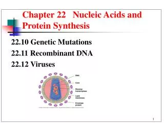
NOTE – DNA replication happens simultaneously at many points along the molecule. It does NOT begin at 1 end and proceed to the other. • NOTE – Replication occurs in OPPOSITE directions on the 2 strands. • DNA Replication is very accurate: - Approximately 1 error in 10,000 paired nucleotides occurs. - DNA proofreading and repair enzymes cuts the error rate to 1 per 1 Billion nucleotides. • Proofreading and Repair Enzymes – Scan DNA for errors, Chemically “snip” them out and “glue” in the correct sequences. • Mutation – a change (error) in the nucleotide sequence….may have no effect or may have serious consequences. Caused by a variety of agents including chemicals, radiation, UV light from sun.
RNA • RNA = Ribonucleic Acid – SINGLE stranded. It moves genetic information from the nucleus to the site for protein synthesis in the cytosol. • RNA is similar to DNA but: - It is SINGLE STRANDED. - The sugar is RIBOSE instead of Deoxyribose. - URACIL replaces thymine.
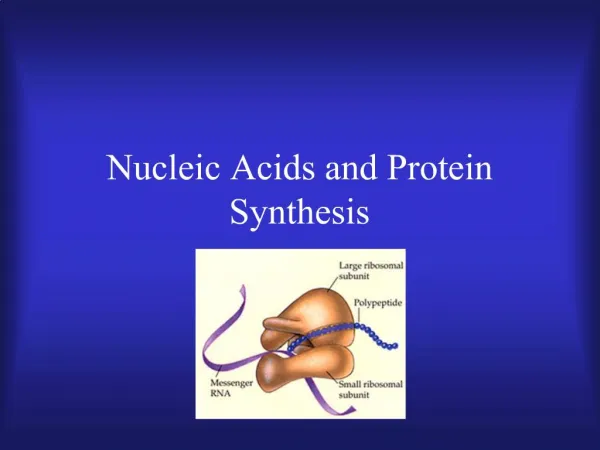
3 Types of RNA:(Each has a different job) • Messenger RNA – (mRNA) – nucleotides are in a single uncoiled chain. It carries genetic information from the DNA in the nucleus to the cytosol. • Transfer RNA (tRNA) – single chain folded into a cloverleaf shape. Binds to specific amino acids. See fig. 10-8 on pg. 194. • Ribosomal RNA – (rRNA) – most abundant form. Its structure is “globular”. It makes up the ribosomes where proteins are put together.
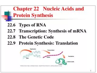
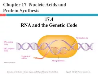
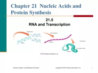
TRANSCRIPTION • Transcription – process by which genetic information (genetic “code”) is copied from the DNA to RNA. OCCURS IN THE NUCLEUS. Read pages 191 – 192. Continued on next slide………..
Steps of Transcription: • RNA Polymerase = transcription enzyme – starts transcription by binding to “PROMOTER REGIONS” ( these regions have lots of A-T base pairs) on the DNA. • Promoter Region – marks the beginning of the DNA portion that will be transcribed • RNA polymerase attaches to 1 strand of unzipped DNA and begins pasting together complementary RNA nucleotides to form a strip of RNA. NOTE – Base pairing rules are the same as in DNA replication, EXCEPT URACIL REPLACES THYMINE!! • Transcription continues until the RNA polymerase reaches a DNA region called the “TERMINATION SIGNAL” = specific sequence of nucleotides that marks the end of a gene or genes. • NOTE – All 3 types of RNA (mRNA, tRNA, and rRNA) are transcribed this way.
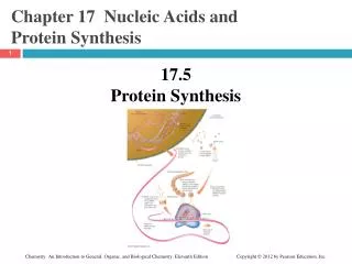
Protein Synthesis • Protein Synthesis = production of proteins. NOTE – all 3 types of RNA will be at work here.
Protein Structure & Composition: Key Points • Proteins are polymers made up polypeptide chains (chains of amino acids – “aa” from here on out). • There are 20 different aa’s that make up proteins. • Proteins may consist of 100’s or 1,000’s of the 20 different aa’s arranged in a particular sequence. • The aa sequence determine how a polypeptide will bend and twist to form the 3-D structure of a protein. • The 3-D structure determines how a protein will function.
Genetic Code – the link between the nucleotide sequence in DNA and the aa sequence in proteins. • Codon – a combination of 3 nucleotides in mRNA. Each codon codes for a specific aa.See table 10-1 on pg. 194. • Facts About the Genetic Code: - several codons code for each aa. - Start Codon = AUG – engages a ribosome to start translation (also codes for the aa –Methionine). -Stop Codon - = UAA, UAG, UGA – cause the ribosome to stop translating.
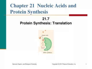
Translation – process of assembling aa chains from information encoded in mRNA. After mRNA is made, it leaves the nucleus and migrates to a ribosome in the cytosol – THIS IS THE SITE FOR PROTEIN SYNTHESIS!! • tRNA – bonds to and transports an aa to the ribosome = “aa taxi cab”. See fig. 10-8 on pg. 194. • Anticodon – sequence of 3 nucleotides that is complementary to pairs with a corresponding mRNA codon. If ACA is the codon, then UGU would be the anticodon.
Ribosome – site for protein assembly. It can float freely in the cytosol or can be attached to the endoplasmic reticulum (ER). It is made up of rRNA and proteins that hold it together. - free floating ribosomes make proteins to be used WITHIN the cell. - ribosomes attached to the ER make proteins to be exported OUT OF THE CELL. • 3 binding sites on a ribosome – (Key to Translation): - 1 site that anchors the mRNA that is going to be translated. - 2 sites that hold the tRNA “taxi cabs”. SEE fig. 10-9 on pg. 195.
Steps of Translation =(Protein Synthesis)See fig. 10-9, pg. 195 • 1. Ribosome attaches to the start codon (AUG) = methionine on the mRNA transcript. Therefore, the 1st aa in every protein chain is Methionine. • 2. As the ribosome move s along the mRMA, each mRNA codon is sequentially paired with its tRNA anticodon (aa taxi cab). • 3. aa’s are hooked to a growing polypeptide chain In the order specified by mRNA. • 4. As each aa is added, the ribosome moves ahead 3 nucleotides (1 codon) on the mRNA transcript. • 5. Eventually the ribosome reaches a stop codon….this ends translation. • 6. mRNA is released from ribosome and polypeptide chain (protein) is complete.
NOTE : - Several ribosomes may simultaneously translate the same mRNA. As soon as 1 ribosome is out of the way, another follows behind. - Translated polypeptide chain = PRIMARY structure of a protein. As it folds and joins other chains, it becomes the functional structure of a completed protein. - mRNA breaks down rapidly. Its parts are recycled and used in the future.