SGLT1 Protein Localization in Parotid Glands of WKY and SHR Rats
This image depicts the immunolocalization of SGLT1 protein in ductal cells of the parotid glands of Wistar Kyoto rats (WKY), diabetic WKY (WKY-D), spontaneously hypertensive rats (SHR), and diabetic SHR (SHR-D). The images show varying intensities of SGLT1 protein in different rat groups.

SGLT1 Protein Localization in Parotid Glands of WKY and SHR Rats
E N D
Presentation Transcript
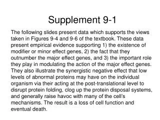
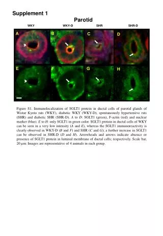
Supplement 1 Parotid WKY WKY-D SHR SHR-D B A C D E F G H Figure S1. Immunolocalization of SGLT1 protein in ductal cells of parotid glands of Wistar Kyoto rats (WKY), diabetic WKY (WKY-D), spontaneously hypertensive rats (SHR) and diabetic SHR (SHR-D). A to D: SGLT1 (green), F-actin (red) and nuclear marker (blue). E to H: only SGLT1 in green color. SGLT1 protein in ductal cells of WKY can be seen in a very low intensity (A and E), whereas the SGLT1 immunoreactivity is clearly observed in WKY-D (B and F) and SHR (C and G); a further increase in SGLT1 can be observed in SHR-D (D and H). Arrowheads and arrows indicate absence or presence of SGLT1 protein in luminal membrane of ductal cells; respectively. Scale bar, 20 µm. Images are representative of 4 animals in each group.