Download

1 / 75
820 likes | 1.23k Vues
Chapter 3: Cells – The Living Units. Cellular Basis of Life. Robert Hooke – late 1600s – observed plant cells 1830s Schleiden & Schwann – concluded all living things are composed of cells Rudolf Virchow – found – all cells come from preexisting cells. 1800s- Cell Theory.

E N D
Cellular Basis of Life • Robert Hooke – late 1600s – observed plant cells • 1830s Schleiden & Schwann – concluded all living things are composed of cells • Rudolf Virchow – found – all cells come from preexisting cells
1800s- Cell Theory • Cell is the basic structural and functional unit of living organisms • Activity of organism – depends on individual and collective activities of cells • Principal of complementary structure and function – biochemical activities of cells are dictated by relative numbers of subcellular structures • Continuity of life from one generation to another has a cellular basis
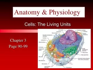
Human Body • Trillions of cells • 200 different cell types • Size ranges from 2 µm to 1 m • Structural unit of all living things • All cells composed of C, N, O, and other trace elements • All cells have same basic parts (generalized or composite cell)
Human Cells • All have 3 main parts • Plasma membrane – outer boundary • Cytoplasm – intracellular fluid packed with organelles • Nucleus – controls cellular activates
Plasma Membrane • Also called the cell membrane • Flexible • Defines the extent of cell • Separates intracellular fluid from extracellular fluid
Fluid Mosaic Model • Thin (7-10 nm) structure • Composed of double layer – bilayer of lipid molecules with protein molecules dispersed in it
Membrane Lipids • Phospholipids – • Polar head – hydrophilic – “water loving” • Non-polar tail – hydrophobic – “water fearing” • Polar heads are attracted to water, lie on inner and out surfaces • Sandwich like structure – heads on outside, tails on inside • Majority of tails – unsaturated – have kinks and increase membrane fluidity
Membrane Lipids • Glycoproteins - lipids with sugars attached • Only on outer membrane surface • 5 % of total membrane • Cholesterol – 20 % of total membrane • Polar region hydroxyl group • Nonpolar region – fused ring • Stabilizes membrane • Increases mobility of phospholipids • Lipid Rafts – 20 % of outer membrane • Assemblies of saturated phospholipids • Packed tightly together • Platforms for receptor molecules and cell signaling
Membrane Proteins • Make up approximately 50 % of membrane • Responsible for specialized function • 2 kinds of proteins • Integral proteins • Peripheral proteins
Membrane Proteins – Integral Protiens • Inserted into membrane • Some stick out only on one side • Others span entire membrane – transmembrane proteins • Hydrophobic and hydrophilic regions • Most involved in transport • Form pores or channels • Others – carriers – bind substances and move them into membrane • Also receptors – relay messages to interior of cell
Membrane Proteins – Peripheral Proteins • Not embedded in membrane • Attached loosely to integral proteins • Include filaments that support membrane • Some are enzymes • Others – motor proteins – change cell shape during division and cell contraction • Glycocaylx – “sugar coating” sugars attached to proteins • Highly specific biological markers – allow cells to recognize each other
Membrane Junctions • 3 factors that bind cells together • Glycoproteins in glycocaylx acts as adhesive • Wavy contours of membrane of adjacent cells fit together – tongue and groove • Specialized membrane junctions a. tight junction – integral proteins of adjacent cells fuse together, prevent molecules from passing b. desmosomes – anchoring junctions, mechanical couplings c. gap junctions – nexus cells connected by hallow cylinders – connections (transmembrane proteins), present in electrically excitable tissue
Membrane Transport • Extracellular fluid – interstitial fluid • Rich, nutritious “soup” • Amino acids, sugars, fatty acids, vitamins, hormones, salts, wastes, etc. • Plasma Membrane – selectively permeable • Allows some substances to pass, but NOT others • Passive Processes – substances cross without energy input • Active Process – substances need energy input to cross
Transport - Passive • Diffusion – movement of molecules from an area of high concentration to an area of low concentration • Down the concentration gradient • Kinetic energy of molecules moves them • Speed of diffusion – influenced by: size – smaller the molecules are the faster they move • Molecules move until equilibrium is reached (no net movement) • Plasma membrane – physical barrier – but molecule will diffuse if it is– • Lipid soluble • Small enough to pass through channels • Assisted by carrier
Transport - Passive • Simple Diffusion – nonpolar and lipid soluble substances diffuse directly though membrane • Oxygen, carbon dioxide, fat-soluble vitamins, etc,
Transport - Passive • Facilitated Diffusion – molecules that can not pass through the membrane by themselves • Transported with the help of a protein • Substance can binds the carrier protein in membrane • Substance may also move through water filled channels • Carriers – integral proteins – allow substances to pass through membrane • Channels – transport proteins • Transport water or ions through aqueous channels
Transport - Passive • Osmosis – diffusion of solvent (water) through a selectively permeable membrane • Aquaporins (APQs) water specific channels in the membrane • Moves down the concentration gradient • Also depends on the concentration of solutes • Osmolarity– total concentration of all solute particles in a solution
Transport Passive • Osmosis cont – • Water diffuses until hydrostatic pressure (back pressure exerted by water against the membrane) with in the cell is equal to its osmotic pressure (tendency of water to move into cell by osmosis) • Tonicity – change in shape or tone of cells by altering internal water volume
Solutions • Isotonic – cells with the same concentration of solutes on the inside and outside • Hypertonic – solution has a higher concentration than the inside of the cell • Water moves out, cells shrinks • Hypotonic – solution is more dilute than inside of cell • Water moves in, cell swells
Active Transport Processes • Active Transport – requires protein • Combines specifically and reversibly with transported substances • Solute pumps – move solutes against the concentration gradient • Requires the input of energy • Symbort System – 2 substances transported the same way (both inside or both outside) • Antiport System – 2 substances transported opposite ways (one inside and the other outside)
Primary Active Transport • Hydrolysis of ATP phosphorylation of transport protein • Protein changes shape – pumps solute across membrane • Ex. Na+-K+ pump – Na+K+ ATPase – drives sodium out of cell and potassium in
Secondary Active Transport • Single ATP powered pump indirectly drives secondary active transport • Na moves back into cell, another substance is cotransported with it • Ex. Sugar, amino acids, ions, etc
Vesicular Transport • Fluids containing large particles and macromolecules are transported in membranous sacs – vesicles • Exocytosis– process that ejects substances from interior of cell
Vesicular Transport • Endocytosis– process that moves substances into the cell • Substances moved in by the infolding of the membrane – coated pit – clathrin – protein coating, then vesicle detaches • Phagocytosis – cells engulfs large solid material • Bacteria, debris, etc • Endocytic vesicle – phagosome • Amoeboid motion – flowing of cytoplasm into temporary pseudopods • Pinocytosis – fluid phase endocytosis • Plasma membrane surrounds small volume of fluid containing dissolved material • Receptor mediated endocytosis– plasma membrane binds only certain substances
Vesicular Transport • Other protein coats – • Caleolae – tubular or flask shaped inpocketings of plasma membrane • Coatomer (COP1 & COP2) proteins – vesicular trafficking • Transport substances between organelles
Plasma Membrane – Membrane Potential • Membrane Potential – voltage – electrical potential energy resulting from separation of oppositely charged particles
Plasma Membrane – Membrane Potential • Resting State – resting membrane potential – range -50 - -100mV • Cell said to be polarized • (-) indicates inside is negative compared to the outside • Diffusion – causes ionic imbalances that polarize the membrane, active transport maintains membrane polarization • Ions – K+ and protein – inside cell • Na+ and Cl- outside cell • Membrane somewhat permeable to K+ - leaky channels • Protein anions cannot follow
Plasma Membrane – Membrane Potential • Membrane becomes negative = -90 mV • Na+ also a factor – attracted to cell interior – bring membrane to -70 mV • Active transport – depends on diffusion • More Na+ in, the more is pumped out • Na+/K+ pump – 3 Na out for 2 K in • Electrochemical gradient – electrical and concentration (chemical) forces
Cell-Environment Interactions • Cell Adhesion Molecules (CAMs) • Key role in embryonic development, wound repair, and immunity • Sticky glycoproteins
Cell-Environment Interactions • Functions – • Molecular “Velcro” cells use to anchor themselves to molecules in extracellular space and to each other • The “arms: that migrating cells use to haul themselves past one another • SOS signals – sticking out from blood vessels lining that rally WBC to infected or damaged area • Mechanical sensors – respond to tension at cell surface by stimulating synthesis or degradation of adhesive membrane junction • Transmitters of intracellular signals that direct cell migration, proliferation, and specialization
Roles • Membrane receptors – integral proteins and glycoproteins that serve as binding sites • Cell Signaling – coming together and touching of cells • Cells recognize each other • Essential for normal development and immunity
Roles 2. Chemical Signaling – • Ligands – signaling chemicals, bind to specific plasma membrane receptors • Include – neurotransmitters, hormones, paracrines • G-Protein – linked receptors – exert effects through G-protein • Second messengers are generated and connect plasma membrane events to internal • Ex. Cyclic AMP – Ca2+ • Activates a protein kinase cascade • Nitric oxide (NO) – another messenger – important signaling molecule
Cytoplasm • “cell forming’ material • Cell material between the plasma membrane and the nucleus • 3 major elements – cytosol, organelles, and inclusions
Cytoplasm 1. Cytosol • Viscous semitransparent fluid • Colloid and solution properties • Dissolved (in water) – protein, sugar, salts, and solutes 2. Organelles - • Carry out specific functions – synthesize proteins, package proteins, etc. • Chemical substances not always present 3. Inclusions - • Stored nutrients, lipid droplets, pigment, water containing vacuoles, crystals of various types
Organelles • “little organs” • Nonmembranous – lack membranes • Membranous – with membranes
Mitochondria • Lozenge-shaped membranous organelles • Power plants of cells • 2 membranes – • Outer – smooth and featureless • Inner – folds inward forming cristae • Gel-like substance inside • Food particles – glucose – broken down into water and carbon dioxide by enzymes • Metabolites broken down and oxidized – energy released and captured • Aerobic cellular respiration
Mitochondria • Contain their own DNA, RNA, and ribosomes • Can reproduce themselves • 37 genes – direct synthesis of 1% of proteins needed • Similar to bacteria (purple bacteria phylum) • Believed to have arose from bacteria that evaded plant and animal cells
Ribosomes • Small dark staining granules • Protein and a variety of RNA – ribosomal RNA • Site of protein synthesis • Some float freely • Others attached to rough ER • Free ribosomes – make soluble proteins • Rough ER – make proteins destined for cell membranes or export
Endoplasmic Reticulum (ER) • “network” with in cytoplasm • Intracellular connected tubes and parallel membranes enclosed fluid filled cavities or cisternae • Continuous with the nuclear membrane • Half of cells total membrane • 2 types – smooth and rough
Rough Endoplasmic Reticulum (RER) • Surface covered with ribosomes • Proteins from ribosomes – enter and are modified • “Membrane factory”
Smooth Endoplasmic Reticulum (SER) • Continuous with the rough ER • No role in protein synthesis • Involved in: • Lipid metabolism, cholesterol synthesis, synthesis of lipid components – of lipoproteins • Synthesis of steroid hormones (sex hormones) • Absorption, synthesis, and transport of fats • Detox of drugs, pesticides, and carcinogens • Breakdown of stored glycogen into free glucose - Muscle – also stores calcium
Golgi Apparatus • Stacked and flattened membranous sacs • Traffic director for cellular proteins • Modify, concentrate and package proteins and lipids • Secretory vesicles or granules migrated to plasma membrane and discharge contents
Lysosomes • Inactive digestive enzymes • Cells demolition crew • Digest particles taken in by endocytosis, particularly bacteria, viruses, toxins • Degrade worn-out or nonfunctional organelles • Metabolic functions – glycogen breakdown and release • Breakdown of nonuseful tissues – uterine lining during menstruation • Breaking down of bone to release calcium into blood - Autolysis – lysosome rupture and cell digests itself
Endomembrane System • System of organelles that work together to: • Produce, store, and export biological materials • Degrade potential harmful substances • ER, Golgi, secretory vessels, and lysozomes, also nuclear membrane
Peroxisomes • “peroxide bodies” • Membranous sacs with a variety of powerful enzymes • Oxidases and catalyzes • Oxidase – use molecular oxygen to detoxify harmful substances – alcohol/fermaldehyde • Neutralize dangerous free radicals – highly reactive chemicals with unpaired electrons • Convert to hydrogen peroxide • Numerous in liver and kidney
Cytoskeleton • “cell skeleton” • Elaborate network of rods running through cytosol • 3 parts – microfilaments, intermediate filaments, and microtubules
Cytoskeleton - Microtubules • Elements with largest diameter • Hallow tubes made of spherical protein subunits- tubulins • Organelles attached • Motor proteins – move and reposition organelles